Prof. Yonat is the fourth woman to win the Nobel Prize in Chemistry, and the first woman to win it in the last 45 years. This article was published in the Weizmann Institute magazine. Coming soon: Excerpts from an interview Prof. Yonat gave last week jointly to the Hidan website and Galileo

The 2009 Nobel Prize in Chemistry was awarded on December 10 to Prof. Ada Yonat from the Weizmann Institute of Science. Prof. Yonat is the fourth woman to win the Nobel Prize in Chemistry, and the first woman to win it in the last 45 years. The prize, which she will share with the scientists Thomas Steitz from Yale University, USA, and Venktraman Ramakrishnan from the National Institutes of Health in Cambridge, UK, was given to them for deciphering the spatial structure and understanding the principles of operation of the ribosome, the cell's protein factory. The achievement, made possible thanks to the development of original research methods, may help, among other things, in improving the effectiveness of antibiotic drugs.
Prof. Yonat was born in Jerusalem in 1939, and completed graduate and certified studies at the Hebrew University. She did her doctoral work at the Weizmann Institute of Science, and after post-doctoral research in the USA, she returned to the Weizmann Institute in the early 70s, and joined the faculty of the Faculty of Chemistry - where she established the first (and for about a decade, the only) laboratory in Israel for deciphering protein structures using X-ray crystallography ("X-ray"). Prof. Yonat won many awards and honors, including the Israel Prize for 2002 and the Wolf Prize for 2007.
Protein factories
At the end of the 70s, Prof. Ada Yonat, who was then a young scientist at the Weizmann Institute of Science, decided to challenge one of the key questions regarding the ways in which living cells operate: to decipher the structure and principles of operation of the ribosome, the factory where the instructions encoded in the genetic code are translated, DNA, to create cell proteins. This was the beginning of a long journey that lasted for decades, and encountered - throughout - reactions of disbelief and even ridicule from the scientific community. Her work, which began in a modest laboratory with a modest budget, expanded over the years and encompassed researchers from all over the world under the leadership of Prof. Yonat. This basic research, which began with an attempt to understand one of the principles of nature, led to the understanding of the way in which several antibiotic drugs work, which may help in the development of more advanced and effective antibiotic drugs, as well as help in the fight against bacteria that have developed resistance to antibiotics - a problem defined as one of the central medical challenges of the century the 21st.
Proteins are the main substances that carry out life processes. Their activity is made possible thanks to their spatial structure, which is determined by the sequence of the building blocks that make them up (called amino acids) dictated by the genetic code. The ribosome, which produces the proteins according to the information contained in the genes, itself consists of a large number of proteins and nucleic acids organized into two subunits: large and small. These units exist separately but work together, in harmony, with maximum efficiency and precision in carrying out the complicated process of creating proteins.
The process of the formation of the proteins in the ribosome is one of the most basic and intriguing life processes. Many scientists in different parts of the world tried for a long time to decipher and understand the way the ribosome works, but their success was partial because the spatial structure of the ribosome was not known. to discover the spatial structure of biological molecules and other bodies - whose smallness does not even allow viewing
In an electron microscope - the scientists create crystals from them. They irradiate these crystals with X-rays (x-rays). Measuring the radiation scattered from the crystal may teach about the spatial structure of the molecules that make it up - to the point of being able to create a "map" describing the distribution of electrons in the studied molecule. This technology is called X-ray crystallography. However, when dealing with the ribosome, it is a particularly challenging problem. It is an aggregate (complex) with a very complex structure, incredibly flexible, unstable and lacking internal symmetry, properties that make it very difficult to form crystals from it, or from its subunits. In addition, the use of X-rays often causes the destruction of the crystals. Another difficulty is the separation barrier (resolution): scientists working in this field have never been able to obtain information about the components of the ribosome at a resolution sufficient to allow them to explain its activity - around three angstroms (which are three millionths of a centimeter).
Crystallizer
During experiments that took place in the Department of Structural Biology at the Weizmann Institute of Science and the Ribosome Research Unit at the Max Planck Institute in Germany, Prof. Yonat succeeded - in the early 80s - in creating the first ribosome crystals in the world using a method for ribosome activation previously developed at the Weizmann Institute of Science by Professors Ada Zamir, Ruth Miskin and David Elson. She is also the first to identify actual evidence of the existence of a "tunnel" within the active ribosome. This tunnel is used to protect the proteins that have just been formed in the ribosome, until the stage where they form into a structure that allows them to "protect themselves". In doing so, she developed several techniques that are common today in the field of structural biology in the world. The most well-known and widely used technique is called cryo-crystallography, which means exposing the crystal to a low temperature - minus 185 degrees Celsius - which prevents its disintegration as a result of strong X-ray irradiation. In addition to this, it has developed unique experimental systems for researching the ribosome, such as this one based on the use of ribosomes taken from bacteria that exist in the Dead Sea. These research methods are currently used by many researchers in many parts of the world.
At the end of the 90s of the 20th century, Prof. Ada Yonat also managed to break the separation limit, thanks to new techniques of crystallography, the result of her development. This is how she managed to get an initial "electron density map" of the small subunit of the ribosome. It is this subunit that translates the genetic code encoded in messenger RNA molecules into the information required for protein production. These findings were published in 1999 in the journal "Records of the American Academy of Sciences" (PNAS). Then, in 2000 and 2001, Prof. Yonat published the first and complete decoding of the two subunits from the ribosome of a bacterium, a work defined by the journal Science as one of the ten most important works of that year. These findings were a high point in research that lasted 20 years, but Prof. Yonat's journey to understand the ribosome has only just begun. Equipped with the new insights she obtained about the structure of the ribosome, she set out to understand how understanding the spatial structure allows us to describe its activity and the way in which antibiotic drugs are used to disable it.
The next task of Prof. Yonat and the members of the research group she heads was to understand the stage at which the first contact was made between the messenger RNA molecule and the ribosome. This contact signals the possibility of starting the production process of a protein according to the genetic information. In order to do this, Prof. Yonat and her colleagues injected into the crystal a certain cellular component, which adheres firmly to the ribosome and initiates its action.
How a drug works
Due to the enormous importance of the ribosome to the course of life, it serves as a target for many antibiotic drugs. "The progress we have achieved in the long journey to decipher the structure and way of functioning of the ribosome may, in the future, pave the way for improving the effectiveness of various antibiotic drugs, aimed at inhibiting the ribosomal activity of disease-causing bacteria," said Prof. Yonat at the time, indeed, her next goal was to find out how drugs affect Antibiotics on the activity of the ribosome. For this purpose, she prepared - together with the members of her research group, Dr. Anat Bashan and research student Raz Zrivetz, and scientists from the Max Planck Institute in Germany - crystals of structural units of ribosomes from bacteria to which one of five antibiotic drugs is attached at a time. Then, based on their comparison with the structure of the ribosome, they deciphered the spatial structure of the "pockets" in which the drugs bind. With this method, the scientists were able to distinguish the molecules of the antibiotic drugs when they are adjacent to the activity sites of the ribosome in a way that thwarts its normal operation. As a result of the adhesion, the cell's protein factories are paralyzed, which disrupts the bacteria's protein production. The proteins are the main biochemical components that activate the various life activities, and a disruption in their production processes causes the bacteria to die. These findings were published in 2001 in the scientific journal Nature.
Since then, Prof. Yonat has managed to reveal the working methods of most of the antibiotics in use, and her research in this field continues even today. Prof. Yonat: "All antibiotic drugs stick to the activity sites of the ribosome. This is how they paralyze it, and prevent the formation of new proteins that are essential for the bacterium. The understanding we achieved regarding the way protein is built in the ribosome will allow the development of more effective antibiotic drugs, which can also harm bacteria that have already developed resistance against existing antibiotic drugs."
Another study, carried out in collaboration with scientists Luis Massa from the State University of New York, and Jerome Carla, from the US Naval Research Institute, winner of the 1985 Nobel Prize in Physics, provided detailed data on how the amino acids - the building blocks from which proteins are made - connect to each other. – in the ribosome. Through the use of a method called "quantum crystallography" the scientists were able to observe the process of protein creation as it occurred. And what's next? Very little is known today about the "birth" of the proteins, that is, how they behave while they grow "stone by stone", and shuffle through the ribosomal channel until they exit the cell, and what happens to them when they exit. These questions are now being investigated in Prof. Yonat's laboratory. In addition, the essential role that ribosomes play, as well as the fact that they are found in every living cell - from yeast and bacteria to mammals - and that their active sites are remarkably conserved from an evolutionary point of view, led to the raising of a theory that early forms of ribosomes are the ones that formed the beginning of life on Earth. In other words, these buildings carried the essence of life. How did these ancient ribosomes form? How did they acquire the structure that allows them to produce proteins? How did the sophisticated factories that exist today in all animal cells develop? These questions are currently the focus of Prof. Yonat's research.
The first experiment
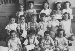
Ada Yonat spent her early childhood in a four-room apartment where her family lived with three other families and their children. The difficult conditions did not suppress her burning curiosity. Already at the age of five, she wanted to explore and understand how the world around her works. She tried to check the height of the balcony in her house using the furniture in the house. She placed a table on a table and on them a chair and a stool on it, and when she reached the end of the tower she fell into the garden and broke her arm. In the kindergarten graduation photo, Ada is seen with her hand in a cast (standing third from the right).
- More of the topic in Hayadan:
- Prof. Yonat: Natural selection also played a role in the probiotic world (before the development of life)
- The ribosome - the key to life at the atomic level - translation of the review explaining why the Nobel Prize was awarded to the discoverers of the ribosome, including Prof. Ada Yonat - part XNUMX
- The ribosome - part XNUMX of the review
- To Part C of the review, in which there is a description of Yonat's discovery about the Dead Sea bacterium
- To the fourth and last part of the review, and further details on Prof. Yonat's discoveries
