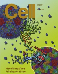
Researchers from the University of California, Los Angeles published in the prestigious journal Cell that they were able to simulate the structure of a virus at such a high resolution that the individual atoms could be distinguished - the first publication ever of imaging biological systems at such a resolution.

"This is the first study in which a structure image was obtained at the atomic level using only this microscopy," said the researcher. "By demonstrating the great usefulness of this microscopy method, we paved the way for extremely diverse biological studies."
Using a standard light microscope, you can get a magnified image of a sample viewed through lenses. Some samples, on the other hand, are too small to scatter visible light and therefore cannot be observed with this method. In order to observe objects whose size is below five hundred nanometers, the scientists are required to use other tools, such as an atomic force microscope, which uses an extremely tiny atomic tip to obtain an image by scanning the surface of the sample, similar to the way blind people read Braille.
Using electron microscopy, another suitable method, a beam of electrons is launched towards the sample, passes through vacuum complexes and is reflected from compressed regions. A digital camera follows the path of electrons passing through the sample to obtain a two-dimensional image of it. By repeating this process at hundreds of varying angles, a computer can obtain a three-dimensional image of the sample with the highest resolution.
Accurate three-dimensional structural reconstruction of biological systems is possible through cryo-electron microscopy because the samples are quickly frozen so that they can be examined in their original environment and because the microscope operates in a vacuum, since electrons move better in such a medium. The article focuses on the structural study of aquareovirus, a non-enveloped virus that causes disease in fish and shellfish, with the aim of better understanding how viruses of this type infect host cells.
"We are very excited about this research group's latest breakthrough," said the dean of the UCLA School of Medicine. "The ability to understand the structure of viruses at the atomic level will pave the way for their utilization in the field of drug delivery and advance certain inventions in the field of curing diseases."
Viruses can be divided into two types: those that have an envelope and those that lack it. Enveloped viruses, which include the influenza and AIDS viruses, are surrounded by an envelope-like membrane that the virus uses to fuse with and infect a host cell. Non-enveloped viruses lack this membrane and instead use a protein to fuse with and infect host cells.
"Through a better understanding of the structures of viruses we hope to develop drugs in three ways," explains the researcher. "If we can understand how the viruses work, we can find small cures or drugs that can prevent the infection; Second, we can make highly stable and harmless virus-like particles as an ingredient in vaccines; And third, we can change their characteristics so that instead of transmitting diseases, they can transmit drugs. Indeed, we are collaborating with doctors and engineers from the university to prepare viruses for gene therapy and drug delivery," explains the lead researcher. "Basically, we hope to take advantage of the millions of years of evolution that have made viruses highly efficient delivery systems."
From the three-dimensional images with high resolution, obtained by cryo-electron microscopy, the research group was able to determine that the virus activates a precursor phase to carry out the infection. In its inactive state the virus has a covering of protective proteins removed during this phase. Once the outer layer is removed, the virus is activated and ready to use a protein called "insertion finger" to infect the cell.
The group's research is a pioneering step for a new era of structural biology in which important biological processes can be understood. The group was able to discover this functionality thanks to the precise structural model created with the help of the unique microscopy. In addition to obtaining a sharp and clear image of the samples, the method allows simulating the samples in their natural environment, so that the structural model is faithful to the original. From a technical perspective, this study also demonstrates the power of this microscopy in creating XNUMXD structures of biological systems without the need for crystal preparation.

6 תגובות
Pretty
Only "distinction" from the word to distinguish (individual atoms) and not "diagnosis" from the word to diagnose (diseases, for example)
Sounds like an amazing achievement indeed. Just out of curiosity, how long do you think it will take from this achievement until any drug that is developed because of this achievement and until we get to see it in the health basket?
I hope that such a microscope will reach Israel.
What's new? I know they do cryo SEM at the Technion too...
Maybe you mean the XNUMXD visualization?
Stunning !
Can you also publish the photos in the magazine?
That is, the sages are of the type that have a shell.