The spread of cancer, or in everyday language - metastasis, leads to up to 90% of deaths related to the disease. Currently, researchers do not have the clinical ability to intervene and effectively stop the spread of cancer cells since many of the steps of this mechanism remain unknown.
After the spread of cancer in the body, the detection and eradication of the spreading malignant cells are essential actions for the survival of the patient. Now researchers report that they have succeeded in developing a new method that allows researchers to mark individual cancer cells in the bloodstream and even track them. This method could help researchers gain better insights into the mechanism by which cancer spreads in the body and develop treatments to stop this spread.
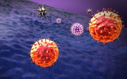
The spread of cancer, or in everyday language - metastasis, leads to up to 90% of deaths related to the disease. Currently, researchers do not have the clinical ability to intervene and effectively stop the spread of cancer cells since many of the steps of this mechanism remain unknown. It is known that cancer cells go through many stages, including invasion of nearby healthy tissues, penetration of the lymphatic system or blood circulation, their spread to other parts of the body, invasion of additional tissues and proliferation in locations far from the source of the tumor.
Now, a new approach developed by Dr. Ekaterina Galanzha from the University of Arkansas allows the marking and tracking of individual cancer cells moving within the body, which will help researchers trace the paths of individual cells from the point of origin to the point of spread. The method is based on taggable luminous proteins that change their color in response to light. When the first laser beam hits the spreading cancer cells, they glow green. A second laser, using a different wavelength, causes the cells to glow red. In order to mark individual cells, the researchers use an extremely narrow violet laser beam aimed at tiny blood vessels.
The reflected radiation from every cancer cell in the body is collected, detected and displayed on a computer screen as real-time signals that allow researchers to count and track individual cells in the bloodstream. "This technology enables the marking of only a single pathological cell from among billions of other healthy blood cells with the help of an extremely fast color change of radiation-sensitive proteins that are inside the cell in response to laser radiation," explains the lead researcher.
With the help of a cancer-infected mouse, the researchers were able to monitor the real-time dynamics of cancer cells flowing in the blood circulation and originating from a primary tumor. They were also able to obtain a picture of the final destinations of the individual cells and of the routes these cells took until they reached the healthy tissues, existing sites of metastases, or the source of the tumor. "Therefore, the approach will be able to provide oncologists with knowledge regarding the way in which it is possible to intervene and stop the spreading cancer cells, thereby preventing the development of metastases," explains the researcher.
This approach could have additional applications in other areas of medicine - for example, tracking bacteria during infections or cells associated with the immune system during the development of autoimmune diseases.
The news about the study

4 תגובות
In addition, in order for the bacteria/cancer cells to express the fluorescent protein, it is necessary to transfect (in the case of bacteria, transformation) a cell that contains the gene for the protein. As of today, I am not aware of the possibility of doing this in vivo... And if there was, we probably could have eliminated the cancer...
What they most likely did here was take a culture of cancer cells, stick them in a dander or something similar and implant it in a mouse... if that's what they did, then there is nothing new here....
On the face of it, this is really not new, the technique has been around for many years....using GFP in its various shades or staining cancer cells are really not something new, as is the attempt to trace the trajectory of the cancer cell...I didn't really understand what they innovated here...
I have no words other than... Wow!!!!!!!!!