TheThe discovery published in the journal Science may lead to the development of innovative technology to treat parasites in the human body and in agriculture

The Technion researchers discovered a family of genes capable of fusion between two mammalian cells, as well as fusion between a virus and a mammalian cell. This discovery, published in the prestigious scientific journal "Science", may have far-reaching consequences in the development of innovative technology to treat parasites in the human body and in agriculture (plants and cattle).
The ability of two or more cells to fuse into one cell is essential for the initial creation of the embryo (fusion between sperm and egg) and for the development of tissues in the body such as the skeleton, muscles and placenta. Fusion between cells is also a crucial step in the infection of the body's cells by enveloped viruses (such as the influenza virus and the immunodeficiency virus - HIV). Despite the importance of the fusion process for human health and reproduction, the mechanism by which cells fuse is unknown.
In the laboratory of Professor Binyamin Podbilevich, from the Faculty of Biology at the Technion, this universal process is studied in an attempt to discover the identity of proteins that mediate the process and the mechanism of their actions at the molecular level. In the laboratory, a nematode (worm) of the type C. elegans is used as a research tool in many laboratories around the world.
In 2007, a family of genes in the worm, which encodes FF (Fusion Family) proteins, was identified for the first time in Professor Podbilevich's laboratory. In a study that has just been published, doctoral student Uri Avinoam discovered that this family of genes also exists in other organisms and that if you remove the FF proteins from different organisms and express them in mammalian cells, they cause fusion between these cells. "At first we thought we had discovered a gene family found only in nematodes (worms)," say the researchers. "We were surprised when we discovered that the genes are also found in other organisms, which makes them, or similar ones, leading candidates to be responsible for the fusion process between cells in the animal world."
In the future, this discovery will make it possible to understand how cells fuse together in the human body and to treat diseases resulting from defects in the fusion process that may cause serious problems in the skeletal and muscular system, and difficulties in fertilization and pregnancy.
Moreover, the Technion researchers were able to express the proteins from the worm on the envelope of a VSV-like virus (Vesicular Stomatitis Virus that cannot reproduce and enters the cell once ("sterile virus"). on top of their envelope. The Technion researchers used the "neutered virus", replacing its envelope protein with a protein from the worm, which allowed it to enter the cell in a one-time fashion. Thus, they showed that the worm's protein, alone, can be used as a fusion protein between cells and in this case - between the virus envelope to the cell envelope. "In order for the fusion to take place," the researchers explain, "the same protein must also be present in the target cell. Since parasitic nematodes have the protein, we can target them with treatment specifically for each parasite."
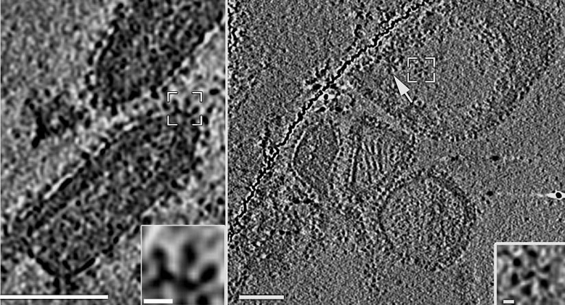
In the study, electron microscopy and tomography were used, with which the researchers photographed the protein on the surface of the "neutered virus" and saw that it forms flower-like structures (in the photograph). In the research, which was done in collaboration with Professor Judith White from the University of Virginia in the USA, Professor Kay Grünewald from the University of Oxford in England and Professor Daganit Danino from the Faculty of Biotechnology at the Technion, master's student Karen Friedman and researcher Clary Valensi also participated.
