Researchers from Tel Aviv University led the international project that may change the map of brain research and medicine

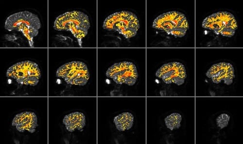
Researchers from Tel Aviv University led a European consortium of 12 groups, which for the first time built a complete atlas of microstructures in the white matter of the human brain. The project, which may change the map of brain research and medicine in the coming years, relied on MRI technology, and was supported by 2.5 million euros from the European Union, in the 'future technologies' category. The partners in the research - known as CONNECT - operate in leading research centers in Israel, England, Germany, France, Denmark, Switzerland and Italy. On Friday, October 19.10.12, XNUMX, at the end of three years of research, they will gather in Paris and announce the successful completion of the project.
Prof. Yaniv Assaf from the Department of Neurobiology at Tel Aviv University, who initiated the international research and served as the consortium coordinator, explains: "Today, medical teams and research groups use an old atlas of the brain, based on the brain of one person, who donated his body to science. The new atlas, however, is based on MRI samples of many subjects, and is even presented in XNUMXD. Therefore, thanks to the research method and scope, it is much more detailed and accurate. In fact, the new atlas allows us to examine the brain in a way that was previously only possible with a microscope."
The first innovation in the atlas lies in the mapping of the white matter - which consists of nerve fibers that transmit information - in the brains of living people, and not in a post-mortem examination. The breakthrough was made possible thanks to MRI technology, which is able to create an image of the living brain in a non-invasive procedure. Second, the new atlas is not satisfied with information from one brain, but relies on imaging the brains of 120 healthy subjects, aged 35-25 years. The data, which was compiled and processed with advanced image processing techniques, gives a broad and accurate picture of the normal brain structure.
The new atlas shows tiny microstructures all over the brain in an orderly and coded manner, which is also suitable for a user who is not an expert in brain research, such as a doctor or a researcher from another field. The images represent various parameters, which were collected and measured using the MRI, such as the thickness of the fibers and their density in the different areas. They are intended to serve as a standard of a healthy brain, to which imaging from the brains of future patients and subjects can be compared.
In addition to its medical contribution, the project is expected to accelerate and significantly advance the study of the white matter in the brain, its activity and functions. "Until today, neuroscientists have mainly focused on what is known as the 'gray matter'," says Prof Assaf. "They avoided investigating the 'white matter', which makes up about half of the volume of the brain, mainly because they lacked effective research methods. The MRI method we developed will allow researchers for the first time to observe what is happening in the nerve fibers in the living brain, and will open up a world of new possibilities for them." Among other things, Prof. Assaf and his colleagues intend to use technology to examine the dynamism and change over time of the microstructures in the white matter. So for example, they will look for the fingerprint that leaves a cognitive process, such as learning a new subject, in the fibers of the brain. Another direction of research is the characterization and understanding of the changes caused by various diseases - such as Alzheimer's or schizophrenia - in the human brain, with the aim of developing effective and reliable diagnostic methods.

3 תגובות
It is very interesting where science has managed to reach, the wonders of technology. Good luck
Hope they come of age for this wonderful thing which is basically the main computer of all creation. Successfully!