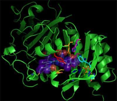
Researchers from Rice University discovered that cutting a certain fluorescent protein and using it as a marker is convenient for examining the activity of living cells

Half protein is better than nothing, and in our case it is even better than whole protein. Researchers from Rice University have discovered that cutting a certain fluorescent protein and using it as a marker is convenient for examining the activity of living cells, especially in understanding the way in which they utilize iron-sulfide accumulations.
Iron and sulfur in the right amounts are essential for good health. They are found in the food people eat and in the vitamins they consume every day, but too much or too little in the cells can cause serious health problems.
Iron-sulfur clusters are compounds with at least four atoms. They are produced and regulated in living cells by proteins and their role is a relatively new field of research for researchers interested in understanding diseases caused by defects in proteins (Friedreich's ataxia, sideroblastic anemia and myopathy). However, until now there was no way to observe these metallic aggregates in living cells.
Researcher Jonathan Silberg, a professor of biochemistry and cell biology at Rice University, has studied the mysteries of these furs for years. He found a way to see how they work in living cells. The researcher and his team published an article in the scientific journal Chemistry & Biology describing the details of the new method, which enables the imaging of the aggregates by attaching them to a mediator fluorescent protein segment.
The mediator is a human protein known as GRX2, an oxidizing enzyme of the glutaredoxin type, which helps cells deal with oxidative damage caused by other proteins in the cell. Its activity can be "turned off" in vitro by linking it to an iron-sulfur pool. The research team has previously demonstrated that this protein will still bind to iron-sulfur clusters even when labeled with a green fluorescent protein; Although the system is useful for in vitro tests, the protein's fluorescence was not strong enough to be seen in live cells.
However, attaching segments of a yellow fluorescent protein, known as "Venus", to monomers of the GRX2 protein worked quite well. When they were injected into living cells, the labeled monomers "found" and used the iron-sulfur clusters as a sort of bridge to link them together. As a result, the segments of the yellow-radiating protein are close enough to each other to radiate strongly enough to be seen under a microscope. The new proteins can be used to diagnose cells to detect signs of diseases caused by irregularities in the concentrations of the aggregates.
"That's why I'm so excited. This is a screen that will make it possible to activate basic biology that no one is able to activate today," the researcher says. "And this system has a high potential to help us find effective treatments against diseases."
The lead researcher says that iron and sulfur were present in the primordial "soup" of the earth even before there was oxygen. "When life developed, the atmosphere on Earth was al-aeronic (lacking oxygen) and iron and sulfur were abundant. It is easy to prepare these metallic aggregates, and therefore, it is easy to imagine how, through simple chemistry and an abundance of these reactants, proteins will evolve and use these aggregates to carry out many chemical processes.
"Then organisms that carry out photosynthesis evolved and began to consume oxygen. Iron is very easy to oxidize, so the appropriate organisms have developed this whole mechanism to protect it and repair it, if necessary. This is exactly the mechanism we are investigating."
The ability to trace these aggregates in living cells is a breakthrough of great interest to the American Heart Association, which funded part of this research. "They gave us funding so that we could develop more tools," notes the lead researcher. "They are interested in understanding the disease Friedreich's ataxia (which can lead to heart disease), but they are also interested in knowing if we can develop additional ways to image other proteins attached to metal clusters."
In this study, he explains, "we actually answered a basic biological question - do proteins of the glutaredoxin type join together in living cells by metal clusters. No one to date has shown this in living human cells. We succeeded in that."
A high priority now exists in the research laboratory for tuning the new tools, however, in the long term, the researcher points out, he recognizes in the new method a potential for examining the roots of aging itself. Abnormal amounts of iron in the human body are toxic, and since oxidation seems to be a major cause of aging, studies of oxidation processes tend to attract a lot of interest.
"Will people age faster because their iron-sulfur pool collection is different? In my opinion, the answer to this question is decades away, but it is a very interesting question - how do small changes in oxidative stress affect aging?
"This is more tempting today after a direct link between defects in the collection of iron-sulfur clusters and the stability of the nuclear genome has been proven," he says. "It's no longer, 'Oh, mitochondrial oxidative stress is somehow related to mutations in the cell nucleus.' There is evidence that defects in iron-sulfur clusters in the mitochondria may be this connection."
