The brain consumes a huge amount of energy - about twenty percent of the body's total energy. Etti Ben-Simon and Michal Gruberger, the Center for Brain Functions, Tel Aviv University and the Tel Aviv Medical Center, researched and found what happens in the brain during rest
The brain consumes a huge amount of energy - about twenty percent of the body's total energy. Current methods of functional imaging indicate that only a few percent of this huge energy budget participates in the performance of specific tasks. What does the brain do with all the remaining energy? What is that "dark energy" of the brain? Welcome to the new field of studying the brain's resting states.
In the mid-fifties of the last century, Louis Sokoloff connected a young college student to a device that checks oxygen consumption in the brain. The American doctor let his subject rest and then had him solve complex mathematical exercises. Sokolov expected a higher oxygen consumption during the mathematical activity, and was surprised when there was no real difference between the brain activity at rest and that during a complex task. In retrospect it can be said that this was probably the first study of relaxation in neuroscience. Today, many findings indicate that the brain's activity is constantly high even if we do not have a specific task to perform, and that the brain's energy consumption is constantly high.
The brain uses 20% of our total oxygen consumption, an average consumption for both cognitive activity states and guiding states (even though it only weighs about 2% of the body's weight). Today it is known that most of this energy is used for internal conversations of the various brain networks with themselves, even when we rest and do nothing in appearance. This intriguing finding was led to by two important discoveries in brain research that miraculously merged in the last decade. In this article we will try to review these discoveries and their meaning regarding brain activity.
The first discovery: the default network
New findings in brain research point to the existence of a network of areas that respond mainly to states of reflection and rest. This network was named The Default Network because it is active by default when the brain is not required to do anything else. This network is always active in the background of every activity we do and there are studies that indicate its connection to the perception of the self, since the perception of the self does not rest for a moment either.
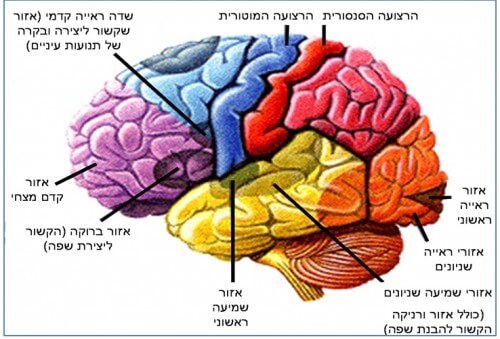
When functional imaging studies of the brain began, for example using PET and fMRI (see an explanation of these methods in Appendix A at the end of the article), most of them focused on brain activity while performing tasks. The hypothesis that advanced this type of research was that without a specific task, brain activity be random and scattered just like white noise (the "snow" when our television is not tuned). This method has yielded many fruits and thanks to it we know how to map many of the brain's functions over specific areas or dedicated networks, such as the parts of the brain active in vision, hearing, decoding language and more As can be seen from picture #1:
With this method, you "record" brain activity during a task (for example, viewing a list of words) and then "record" brain activity during rest (for example, when you stare at a point on the monitor). To see the brain activity during the task only, and so that we can filter the "white noise" that we assumed exists in the brain activity, we subtract the resting image from the task image. In this way we see which areas were active, or in which areas the activity decreased, at the time of the task. An example of this method can be seen in picture #2:

This method represented a broader view of brain activity. The basis of this view was the idea that the brain, similar to a computer, mainly responds to the activity of the environment on it and as long as we do not "ask" it to perform a certain task, such as opening a Word document or sending an email, it will be in a kind of standby mode and we will not see any special or organized activity in it. But all this was about to change in the early XNUMXs.
In 2001, a group of researchers led by Prof. Marcus Raichle from the University of Washington published an article in the scientific journal PNAS in which they noted the existence of a network of areas in the brain that actually shows a decrease in neural activity, rather than an increase, while performing a wide variety of tasks. This network consistently responds with a decrease in activity, Regardless of the type of task given to the subjects, whether it is viewing words, memorizing a list of objects, or another task. If the level of neural activity in this network decreases during a task, it means that during rest (the absence of a task) it is working vigorously! The researchers called this network the default network because They concluded that this is the default activity of the brain as soon as we do not give it a defined task but rather "let it go to its own devices". It is interesting to note that once the network was named, many researchers in the field of functional imaging of the brain noted that they also saw a decrease in the activity of these areas while performing tasks, but not A breakdown of the default network areas can be seen in image #3:
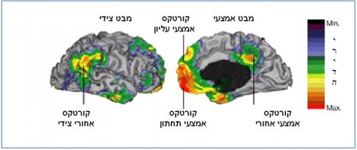
Adapted with permission from: SEARCHING FOR A BASELINE: FUNCTIONAL IMAGING AND THE RESTING HUMAN BRAIN, D. Gusnard & ME Raichle PNAS 2001
.
The team of researchers from Washington led by Reikel wondered: Why do we have a neural network that seems to function while we are resting? Which led to an even bigger question: what does the brain actually do when we stop working?
We invite you to think for a moment: what usually happens when you are not busy with something in particular? The answer is very simple: you are thinking, maybe pondering about your life, the activities you want to do or the experiences you had yesterday. This type of thinking is defined as spontaneous thinking, one that is not necessarily related to the environment. This phenomenon is such an obvious and essential part of our lives that we tend to take it for granted. People who for one reason or another, for example a neurological injury such as a stroke, lost for a short period the ability to manage their thoughts (or in other words - their stream of consciousness) report a very strange experience of disconnection, a lack of connection to themselves. It is so hard to stop "flowing" with thoughts, that even holding your breath seems an easier task to do!
Spontaneous thinking: "Discovery of a new continent"
Spontaneous thinking (in English: mind-wandering/stimulus independent thought) is defined as reflection that is not necessarily related to external stimuli. It is possible to bring a familiar example to drive home where we are pondering, grasping shingles, and suddenly find ourselves near the house. This kind of kind of daydreaming is actually turning our attention to our inner world when in the outer world we go into a kind of automatic state. A familiar activity that does not require concentration will usually lead to spontaneous thinking, as well as the obvious cases where there is no task at all to perform.
Indeed, these are the classic cases in which we see the activity of the default network. The activity takes place whether the subjects are at rest, flowing with their thoughts, or whether we ask them to perform a task that is familiar to them. Studies even indicate a direct relationship between default network activity and the ease with which one can perform the task (and actually escape to contemplation): the easier the task, the more network activity there will be. Spontaneous thinking is not an easy subject for scientific research because by its very nature it is very subjective and therefore difficult to measure externally. By using questionnaires and reports from subjects it is possible to quantify how much the subjects were "in their own world" and these reports do indicate a close connection to the activity of the default network. The investigation of the default network and its connection to spontaneous thinking has led in recent years to many brain studies around the world, in which the stream of consciousness of the subjects was studied in resting states.
It is surprising to discover how such a well-known and essential topic in our lives did not receive proper research attention until the discovery of the Internet. Since the publication of the article and the labeling of the network as the default network, many studies conducted using different imaging methods have established its position and today it is considered one of the well-known functional networks in the brain, just like the attention network (related to the ability to pay attention) or the motor network (related to the planning and control of movement). Research has revealed the existence of this network in animals as well, a fact that indicates that it is a functional network of evolutionary significance, which is not necessarily unique to humans. In studies conducted on patients with mental disorders (such as Alzheimer's, schizophrenia or autism), differences between patients and healthy people were discovered in the degree to which different parts of this network communicate with each other, evidence of the importance of the network's activity for a normal mental life. In studies done on children, it became clear that the default network does not finish its development at young ages, a fact that may imply the importance of experience in shaping the connectivity of the network. Giulio Tononi, one of the leading neuroscientists in the world, described the discovery of a new functional network at such an advanced stage of brain research as "the discovery of a new continent". If there is a network of regions that work together at rest, and which has been preserved in evolution - what exactly does it do?
There are several theories today regarding the role of the default network. The first theory is related to spontaneous thinking and suggests that the network acts as a kind of simulator, simulating life, so that it uses past memories and self-reflections to simulate life and to generate new insights about the world. This theory is remarkably suitable for the various parts of the network. The default network includes extensive areas in the midline region of the brain, an area that buffers between the two halves of the cerebrum, the hemispheres. The frontal area in the midline of the brain (medial pre frontal cortex) is known as an area that is active in the evaluation of stimuli from a personal perspective (for example, does this picture please me or not?) as well as in tasks related to thinking about the self: for example, thinking about character traits that correspond to my self-concept, Like am I a responsible type. Areas in the parietal lobe and the lateral lobe of the network are related to the retrieval of memories, and especially the hippocampus, which is active during the retrieval of new memories or their creation. These two areas are active together in the daydreaming mechanism, with the parietal-lateral areas providing memories of the past and the middle areas evaluating and examining these events from a personal point of view. An extension of this theory suggests that the role of the default network is to filter which memories to retain and which not by assessing their relevance to the self.
A second theory relates to the role of the default network in internal attention. Inner attention refers to a mental activity in which the person is not attentive to his surroundings but to his inner world. In contrast, external attention is directed towards the environment and with its help we pay attention to external events. For example, watching a movie will be defined as an activity that requires external attention compared to daydreaming which is a distinct activity of internal attention. From brain imaging studies it became clear that there is a spontaneous exchange between activity in the default network, which as mentioned represents the activity of internal attention, to activity in the network of external attention while the subjects are at rest. That is to say, the correlation between the activity of the default network and the activity of the external attention network is negative, so that when one increases the other decreases and vice versa. This fact has led researchers to hypothesize that the brain constantly checks the state of the internal environment with the help of a default network and the external with the help of the external attention network, and that these checks are done spontaneously and automatically.
This theory has already led to thoughts about the existence of a third network whose role is to balance or direct the activity between the two attention networks. One can easily think of situations where this balance is violated and we are too inclined to pay attention internally or externally. A well-known example is an increasing tendency to external attention in cases of attention deficit hyperactivity disorder, where at least activity is observed in the internal attention network, which is the default network. In studies done on people with attention deficit hyperactivity disorder, lower connectivity in their network was indeed discovered compared to its connectivity in healthy people. There is a need for additional rest studies using different imaging methods to decide about the primary function of the network and about its importance for the proper existence of the self-concept. It is possible that the activity of the default network is diverse, so that all theories faithfully represent its various functions, and a future model could combine them all for a comprehensive and satisfactory explanation of the importance and activity of this network.
The second discovery: the "plots" of the brain while resting
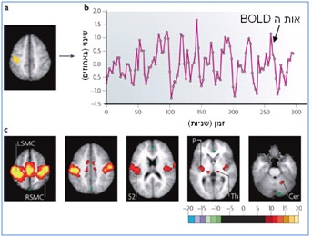
The discovery of the default network and delving into the study of the brain during rest led as mentioned to the intriguing question: what does the brain actually do when we do not present it with a task? This question gave rise to a new and interesting field of research, the study of the brain's resting state networks. It turns out that the default network is just one of several functional brain networks that exhibit spontaneous activity during rest. When we examined the brain without the task, they saw that the BOLD signal obtained from the fMRI device changes spontaneously even if the subjects are lying with their eyes closed and they are not performing any task. These changes are relatively slow, about one cycle every ten seconds, and they work in synchronization between brain areas that are functionally related. For example, if we focus on a brain signal that changes spontaneously (all brain signals change spontaneously during rest and there is no need to look for a specific signal) in a certain motor area in the front of the brain and check which other areas of the brain change in the same way, then we will identify the entire motor network in the brain: in other words, all The areas related to the execution or planning of movement. This research method is called functional connectivity, and you can read more about it in Appendix B at the end of the article. With this method, all known functional networks in the brain were tested and it turned out that they are all interconnected during rest (including the default network) as you can see in picture #4:

Adapted with permission from: Two views of Brain Function, ME Raichle TICN 2010
This means that the brain areas that make up each of these networks talk to each other constantly and spontaneously without any assignment. It is important to emphasize that even though this is a spontaneous activity, there is coordination between the different networks in the brain. From this we can understand that what we thought was random noise is not really random, but rather reflects the functional wiring of the various brain networks. Why does the brain maintain such organized and vital activity during rest?
This question is still being researched but findings from animal studies raise some interesting possibilities for the role of spontaneous activity in the brain. We will mention two prominent studies that may hint at these roles. In a 2006 study published in the science weekly Nature, the activity of cells in the hippocampus (the area responsible, as mentioned, for recording memories) of mice was tested while they were learning to navigate a maze. The researchers continued to record the activity of the neurons even when the mice were resting, and apparently did not do any activity. The researchers showed that while resting, the hippocampus cells of the mice continued to fire in the reverse order of the learning sequence that the mice learned a few minutes earlier in the maze. This means that during rest the activity of the cells helped to retain the knowledge learned earlier. This study suggests the possible importance of spontaneous activity during rest for learning processes. A discovery that fits well with the popular perception that it is desirable to take time off from a demanding task to let the "material sink in".
Another possibility for the role of spontaneous activity in the brain comes from a study conducted at the Weizmann Institute of Science and published in the weekly Science in 1996. In this study Prof. Amos Arieli and his colleagues investigated the activity of the visual area in the brain of cats. It is a well-known fact in brain imaging studies that the response to a certain stimulus (for example the response in the visual area of the brain to an image of stripes) will yield a slightly different result each presentation of the stimulus. In order to see the activity characteristic of a stimulus, it is necessary to elicit a large number of responses from that particular area of the brain. Arieli and his colleagues showed that the explanation for the difference in brain responses stems from the spontaneous activity of the brain that preceded the time of the appearance of the stimulus. They showed that if you take into account the brain activity before the stimulus appears, you can accurately predict how the brain will react to the stimulus without needing to average several stimuli. The significance of this research is that the spontaneous activity in the brain may influence and even predict what our response to stimuli in the world will be.
If we take this hypothesis one cautious step further, we can assume that the spontaneous activity may even determine the rule of our behavior when we are faced with stimuli from the environment and determine why we will react differently at different times when the environment presents us with similar challenges. You can think of it a bit like the "mood of the brain" that affects our reactions to the environment. This study suggests that we take into account the spontaneous activity in the brain when we try to predict the brain activity and not lose this valuable information by filtering it as noise.
A functional organization of spontaneous activity in the brain is found in humans and animals even under anesthesia and even in vegetative states ("plant" states), which implies a very basic and essential organization in brain activity that was discovered to us thanks to the new research field of resting states. Since it is possible to examine the brain's activity and its functional connectivity while resting without the requirement to perform tasks, we can study the brain's activity without the need for any cooperation from the patients, and thus it is possible to examine the brain's connectivity even in states of unconsciousness (coma).
Summary
Brain activity during rest is organized both in time (as coordinated activity between the brain networks) and in space (in the existence of the default network). There is well-coordinated activity between the brain's functional networks even without a specific task to perform. This organization in brain activity exists even without awareness, since like In our presence, spontaneous changes in the BOLD signal were found in functional magnetic resonance imaging even under anesthesia. The network that led to this interesting discovery is the default network. This network is active when we daydream and its activity decreases when we need to perform a certain task. The default network and the study of brain activity at rest allow us to glimpse To the secret activity of the brain, to the constant conversations that the brain has with itself. The effects of this spontaneous activity on the way we function in the world, and even on how we are who we are, are fascinating questions for the field of brain research in the coming years.
Thanks:
We would like to thank Prof. Thelma Hendler and Dr. Avraham Tsangan for their support and Nir Lahav for his helpful comments.
Etti Ben Simon and Michal Gruberger are doctoral students at the Center for Brain Functions at the Tel Aviv Medical Center and Tel Aviv University.
for further reading:
• Michael D. Fox & Marcus E. Raichle, Spontaneous fluctuations in brain activity observed with functional magnetic resonance imaging, Nature Review Neuroscience 8, September 2007.
• Marcus E. Raichle, The Brain's Dark Energy, Science 24, November 2006.
• Marcus E. Raichle, Two views of brain function, Trends in Cognitive Sciences 14, April 2010.
• Malia F. Mason et al, Wandering Minds: The Default Network and Stimulus-Independent Thought, Science 19, January 2007.
Appendix A: Functional Magnetic Resonance Imaging (fMRI) BOLD, and Positron Emission Tomography (PET)
The positron emission tomography (PET) device and the functional imaging device using magnetic resonance
(fMRI), are two different devices used for functional imaging of the brain, i.e., for imaging the brain during activity (as opposed to MRI, which allows examining only the structure of the brain). In PET imaging, radioactive isotopes such as oxygen 15 are used, which the subjects breathe or drink (for example, water that contains radioactive oxygen). Since a radioactive atom is unstable, it decays - and in this case it is beta decay - while emitting positrons. The radiation is picked up by the sensors surrounding the subject. From the angle of emission you can tell where the radioactive oxygen was. With this method it is possible to know what the oxygen or glucose consumption of the brain was in different areas and hence to calculate the brain activity in that area. Functional MRI examines the change in the flow of oxygenated blood in the brain. During neural activity in a certain area of the brain there is a demand for more oxygen and therefore an increase in the amount of oxygenated blood in the active area. MRI uses a strong magnetic field and radio frequency transmission to detect the amount of oxygenated blood. Because the hemoglobin molecule in the red blood cells contains iron, it is detectable within the magnetic field. The difference between oxygenated and non-oxygenated blood is possible because the presence of oxygen changes the magnetic properties. The subjects are in an environment of a strong magnetic field that allows us to detect these changes and thus calculate the amount of oxygenated blood in a certain area of the brain. The signal obtained from functional imaging of the brain is called Blood Oxygen Level Dependent (BOLD) and shows the changes in oxygenated blood flow to different areas of the brain.
Appendix B: Functional connectivity
Adapted with permission from: Two views of Brain Function, ME Raichle TICN 2010
The difficulty in studying the resting brain is that there is no task to perform. We usually subtract the task image from the rest image
(baseline) to see the specific brain activity during the task. When interested in researching resting conditions, we do not have a resting image to compare with. To test brain activity while resting (and actually in the absence of a task) we need new research methods. The subtraction method described in the article is not suitable for studying spontaneous activity during rest, because in this method the rest activity is filtered by subtracting it from the task picture (see Theo above). One of the accepted methods for studying brain activity during rest is called functional connectivity. With this method, a specific area of the brain is selected (for example, the area marked in yellow in the figure below) and a BOLD signal is produced from it. In the next step, all other areas of the brain are examined, to see which of them show a similar signal. It is assumed that regions working together will show a similar signal, therefore the regions found will belong to the functional network from which the initial region was taken. For example, if the initial signal was received from an area belonging to the attention network, the functional connectivity method would allow us to identify the entire attention network in the brain by examining the signal in one area of the network. In this way it is possible to get a mapping of all the functional networks in the brain while resting.

15 תגובות
Peace
The article is very interesting for me.. Thanks for sharing
I have been meditating for about 15 years and the "self" is the central issue that undergoes transformation as a result of this process.. I am intrigued to participate in research to see the difference that develops from the very practice of meditation and to see which different networks operate in which states of consciousness when I choose to enter them.. I would be happy if we could cooperate on questions These and, of course, other questions that will arise.
Very interesting article! Thank you!
I agree with what Eyal wrote here. From personal experience and familiarity with attention disorders, our distraction is not necessarily to external stimuli, but also to completely internal stimuli (thoughts that run without control). One of the amazing effects of Ritalin, for me, was the inner peace it created. Suddenly I could listen to what was going on outside without the internal flood of thoughts interfering. Until then I didn't even know it was possible.
Link to an interesting article:
http://www.calcalist.co.il/local/articles/0,7340,L-3572606,00.html
by the way,
It is possible that the division of attention actually helped those animals to pay attention to other factors when eating.
You can read about serious geniuses, most of whom (if not all) suffered from attention deficit disorder, and there are quite a few studies that confirm that this is typical of smart and creative people. Perhaps it is the tireless curiosity and the surrounding stimuli and the feeling that there is an ability to contain it.
I am sure that it is important and essential to be present, but sometimes the mind is not sufficiently challenged by the current "task".
A problem for most of us is that the mind constantly prepares the future and constantly prepares to "be" in a certain pattern "like" this way and that and this, in my opinion, goes against the essence of thought which should be without limitations
Hi Eti, thanks for the reply, waiting for an email from you.
By the way, there is no contradiction between what he wrote and the concept I presented.
You wrote: Energy is a precious resource in nature and the dipole network is one of the biggest energy guzzlers in the brain.
Truth: in self-awareness work, one of the most important goals is to preserve mental energy that is wasted in huge amounts and does not allow the development of higher functions.
Both in her description and from my personal experience, the main factors that consume this energy are: imagination and chatter, negative emotions, unnecessary physical tensions and the uncontrollable grasping of attention on every stimulus that comes.
You wrote: It is very likely that she would not have survived in evolution if she had not played an important role in the survival of humans or other mammals.
Indeed, daydreaming and endless chatter is a characteristic of the civilized man, the man who has already left the framework of natural selection. If we examine tribal people living in nature, it seems that they live mainly in the present, and much less of us are in the imagination.
I don't know how this network works in mammals or other animals, but I'm pretty sure they use it differently than modern humans do. It's enough to look at animals that live in the wild (those that live next to modern man are already a bit distorted). The antelope grinds grass leisurely, it is not alert to danger, but as soon as it hears the slightest rustling it immediately steps away. If her attention was occupied with imagination she would not have heard slight noises like stepping on a branch and would have been hunted. So from an evolutionary point of view it doesn't make that much sense that the role of the network would be a daydream, or at least one that comes at the expense of the present.
Hi Tsur
Thank you for your response, I will certainly be happy to contact you.
Regarding the role of the diplot network, as you probably know energy is a precious resource in nature and the diplot network is one of the biggest energy guzzlers in the brain. It is very likely that it would not have survived in evolution if it had not played an important role in the survival of man or other mammals. Viewing daydreaming as a waste of time at best and a detriment to happiness at worst are not always justified. There are indeed studies that show that spontaneous thinking can cause a deterioration in mood, but there are also studies that indicate the contribution of this state to creativity and problem solving. Therefore, I think of spontaneous thinking as an infrastructure that can be charged in different directions depending on what is chosen. By the way, also a quiet and present state or in other words a constant perception of the self is an important aspect of the diplot network and maybe even before the 'chatter' that follows it.
To me:
First of all, a fascinating and interesting article.
I have noticed that in all theories that try to explain the function of the default network there is a hidden underlying assumption and that is that this network has a certain role (perhaps even evolutionary). The theories try to explain what the role is.
Have you ever thought that perhaps the high activity of this network while resting from chores, actually constitutes a failure or distortion in the proper functioning of the brain, rather than a natural activity that has a role.
I don't know the answer of course, but I hypothesize this as a person who has been engaged in meditation and self-awareness for many years, and one of the basic ideas shared by the teachings dealing with self-awareness is that healing in the summer, and the endless and involuntary internal chatter (the matrix) is a distortion of our nature. According to this view, the natural and normal state of the human mind is to be quiet and present. That is, he is in the present, and does not run away to the past or an imaginary future without us explicitly asking him to do so.
I belong to a group of practitioners who engage in a practice aimed at quieting the mind and producing silence and presence in every moment of everyday life (not only in sitting meditation). You wrote that moderators are welcome, so you are welcome to contact me and maybe we can interest the group in participating in the study.
Best regards,
flint
zur.brener@gmail.com
Great article!! Thank you!!
Absolutely fascinating article.
Note: I actually know that people with ADHD are more inclined towards daydreaming. This very thing of lack of concentration is being in your own world instead of listening and in an uncontrollable way. That is, according to the article above, their default network is too active, or rather there is poor control over the transition between the networks - between internal attention and external attention. I am like that.
An absolute pleasure, thank you!
Thanks for the compliments! Regarding meditation practitioners it is indeed known that they experience fewer reflections. We are just trying to deepen our knowledge on the subject in our current research. (Meditators are welcome :)
Fascinating and profound
Interesting and engrossing article, thank you!
Reflections –
I am interested in whether people who practice meditation are tested. Meditation usually works precisely on these resting networks (if I understood correctly) and their ability to control the current flow of thoughts is better than that of a person from the village. And if we add the section about Prof. Amos Arieli and his colleagues, is it possible that meditation helps to "direct" the activity before the groi and thus the reactions as well as the reaction in its wake.