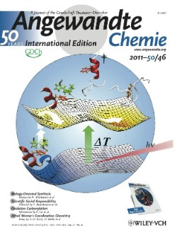
A team of chemists from the University of Pennsylvania succeeded in developing a method in which proteins can be seen to fold "in real time", a method that could lead to a better understanding of the correct and incorrect folding of proteins

The activity of a protein depends on both its composition and its folding form. While deciphering the composition is relatively simple, uncovering the form of folding is a huge challenge and has serious consequences since many diseases break out as a result of improperly folded proteins. Now, a team of chemists from the University of Pennsylvania has succeeded in developing a method in which proteins can be seen to fold "in real time", a method that could lead to a better understanding of the correct and incorrect folding of proteins.
The findings of the study, led by chemistry professor Feng Gai from the University of Pennsylvania, were published in the scientific journal Angewandte Chemie.
"One of the reasons why understanding what happens when proteins fold is problematic is that we don't have a device equivalent to a high-speed camera capable of capturing the process," explains the lead researcher. "If the process was slow, we would be able to collect many "pictures" over time and see the activity of the mechanism. Unfortunately, no device has this capability; The folding happens quickly in the blink of an eye."
The research team used infrared spectroscopy - a method that measures the amount of light absorbed by different parts of the molecule - in order to examine the structure of the protein and how it changes. In this case, the researchers examined a model protein using an infrared laser. In their experiment, the researchers used two lasers to examine the changes in structure as a function of time. The first laser is used to heat the molecule, the step that initiates the structural changes. The second laser functions as a camera that follows the movements of the building blocks of the protein - the amino acids.
"The protein consists of different groups of atoms, when these groups can be considered as springs," explains the researcher. "Each such group has a different frequency at which it oscillates back and forth, a frequency based on the mass of the atom at the end of the group. If the mass is greater, the spring oscillates more slowly. Our "camera" is able to follow the speed of this movement and we can then attribute it to the atoms that make up the protein and how this section of the protein moves."
Even in a simple protein, such as the model protein used, there are many identical bonds and the researchers are required to distinguish between them in order to see which of them moves during the folding of the protein. One of the strategies they used to get around this problem was to use the molecular equivalent of a tracking device. "We used an amino acid that contains a traceable carbon isotopic marker," explains one of the researchers.
With a single carbon atom in the model protein that is slightly heavier than the others, the researchers were able to use its "signature" to infer the position of other atoms during protein folding. In the next step, the researchers can "tune" their laser frequency to match other parts of the protein, so that they can be isolated for their testing.
Similar isotopes can be injected into more complex molecules, which will also allow observing the folding of these using infrared spectroscopy. "This method increases the capacity for structural separation - it allows us to see the moving parts," explains the lead researcher. This ability will allow us, for example, to see exactly how a protein misfolds during a disease.

4 תגובות
"Max" who are you?
And who is Bob?
Boris???
Bob is that you?
If I understand correctly, then I realized that we are slowly starting to see the construction of the secondary structure alpha helix and beta surfaces, in an even deeper way - to see what the incorrect folding looks like and in which part of the polypeptide structure being built the error occurs.
Does the research bring us closer to understanding where and why the protein folds the way it does, and not in a different way? (It means that there are like hundreds of thousands of options for folding, but it folds in a specific way with minimal energy investment)?
Thank you for a to-the-point answer 🙂