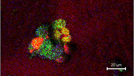
The new method makes it possible to obtain images not only of the borders of the cells but also of certain molecules that are inside the cells or on their surface, as well as their exact location

Researchers from the University of Gothenburg in Sweden have developed a new method for imaging individual cells. The new method makes it possible to obtain images not only of the borders of the cells but also of certain molecules that are inside the cells or on their surface, as well as their exact location.
Individual human cells are extremely small and have a diameter of approximately one hundredth of a millimeter. In light of this, special measuring equipment is needed in order to differentiate between the different parts inside the cell. Researchers often use a microscope that magnifies the image of the cell and shows the outline of its borders, but this device does not provide any information regarding the molecules found inside the cell and on its surface.
"Our new sample holder is full of trapped cells," says researcher Ingela Lanekoff. "In the next step, we quickly freeze the sample to a temperature of minus 196 degrees Celsius, which allows us to obtain images of the various molecules present at the moment of freezing. Using this method we are able to produce images that show us not only the borders of the cells but also the molecules that are inside them, as well as their exact location."
And why are the researchers interested in knowing what the molecules are in a single cell? Because the cell is the smallest living element that exists, and the chemical processes that take place inside it are of great importance to the functioning of the cell in the living body. For example, in our brain there are special cells that are able to communicate with each other using chemical signals. It has already been proven that this essential communication depends on the specific molecules present in the brain cell membrane.
The images of the molecules found in the membrane of individual individual cells allows researchers to measure the changes that take place within them. Together with previous studies, the new findings demonstrate that the rate of communication between the examined cells is affected by a change of less than one percent in the amount of a certain molecule in the membrane. This finding implies that the communication between brain cells is closely dependent on the chemical composition of the membrane of each and every one of the cells. This finding is an important part of the collection that could lead to plausible explanations about the mechanisms underlying human learning and memory.
The research was published in the form of a PhD thesis entitled: "Analysis of Phospholipids in Cell Membranes Using Liquid Chromatography and Mass Spectroscopic Imaging", submitted to the University of Gothenburg, Department of Chemistry.
The news about the study
A link to the doctoral thesis
