In marine fortifications, the building blocks of the minerals are extracted from seawater. In the fetuses of humans and other mammals, the minerals, which come from food, are supplied through the mother's blood.

Imagine an underwater construction site: the floating building blocks are carried into the space where the scaffolding is built. This is how the skeleton and other mineral structures are formed in the embryos of many animals. In marine fortifications, the building blocks of the minerals are extracted from seawater. In the fetuses of humans and other mammals, the minerals, which come from food, are supplied through the mother's blood.
A research group, led by Weizmann Institute of Science scientists, followed the first stages of skeletal construction, starting from the moment of fertilization, in live sea urchin embryos. As recently reported in the Journal of the National Academy of Sciences of the USA (PNAS), they reached surprising insights about this complex process.
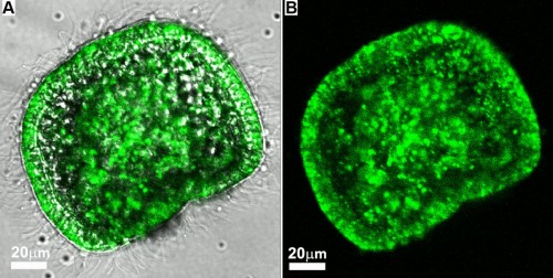
In order to accumulate a sufficient amount of minerals, a sea urchin fetus, whose size is equal to the thickness of a human hair, needs to use all the calcium that contains an amount of water whose volume is hundreds of times greater than its own volume. For decades, scientists believed that the accumulation of calcium extracted from the water and the construction of the skeleton were carried out exclusively by specialized cells of the fetus. But in the new study, in which the scientists followed the development of sea urchin embryos in water containing calcium ions labeled with a green fluorescent dye, they were amazed to find that the entire embryo quickly turned green. It seemed that each of the embryo's cells accumulated calcium ions.

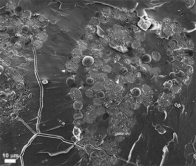
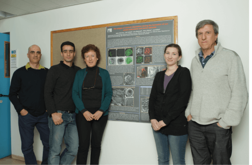
To make sure that the finding was not made by chance during the measurements, the scientists videoed the wide distribution of the calcium using several advanced technologies: in addition to observing live embryos with a light microscope, they studied frozen samples of embryos with a scanning electron microscope. Furthermore, they used state-of-the-art technology developed in Israel, which combines a fluorescent microscope, chemical element mapping, and a scanning electron microscope, which allows the samples to be viewed in normal air instead of a vacuum. The research was carried out by Prof. Leah Addi, Prof. Steven Weiner, and research student Neta Vidavsky, from the Department of Structural Biology at the Institute, together with the Institute's graduate, Dr. Sefi Addi, from B-nano Ltd., Dr. Yulia Mahmid, a graduate of the Institute who works Today in research in Germany, Dr. Eil Shimoni from the Department of Chemical Research Infrastructures at the institute, and David Ben-Ezra and Prof. Moki Spiegel from the company "Exploring Seas and Lakes for Israel" in Eilat.
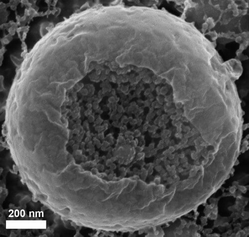
In addition, it was discovered in the research that when calcium carbonate enters the cells of the embryo, it forms granules made of nanospheres. This structure is typical of many amorphous minerals, and is known as an intermediate stage in the construction of skeletal tissue in sea urchins, as Prof. Weiner and Prof. Addi discovered over a decade ago. Now the research has revealed that the grains are kept inside vesicles.
The findings point to new research directions in regards to the formation of the skeleton in a variety of living creatures, including humans. The fact that the entire embryo is mobilized to build the skeleton in the sea urchin suggests that, contrary to popular opinion, even in humans, other cells, besides the specialized cells, called osteoblasts, may be involved in the building of bones and teeth. Furthermore, it is possible that the temporary storage of calcium carbonate in vesicles also exists in other creatures, besides sea urchins.

Scanning electron microscope photograph: A frozen sea urchin embryo containing a large number of intracellular vesicles
The study of these mechanisms is of great importance for a better understanding of biological mineralization processes. This understanding may be essential for future studies on various diseases and structural problems of the bones and teeth.
For previous articles on the subject:
- What can be learned from the sea urchin?
- The sea urchin genome is deciphered
- Bio Engineering - Sholit Nachshon Hayam

