The breakthrough will enable an understanding of how complex networks of brain cells work through the use of "optoint" cultures
Networks of brain cells growing on a growth medium in laboratory plates (two-dimensional cell cultures) are a convenient model for many studies in the field of neuroscience and medicine. Their great advantage is the relative simplicity in which it is possible to test physiological changes in cell activity patterns, resulting from changes in their environment. However, the disadvantage of cell cultures is that they consist of a single layer of cells, and do not include the complex, three-dimensional connectivity of brain cells found in the human body that form complex neural networks.
Previous attempts to develop three-dimensional models for the study of the central nervous system were not very successful, both due to the high complexity of developing a three-dimensional culture simulating the tissue of the brain and due to the challenge of developing methods to study network activity in three dimensions. Technion researchers have now succeeded for the first time in developing three-dimensional cell networks that simulate complex aspects of brain activity, something that will enable an understanding and influence on the physiology of the central nervous system. This is what the prestigious scientific journal Nature Communications publishes.
The editor of the journal notes that three-dimensional neural networks are a promising model of complex neural tissues, which may lead to an understanding of the structure and function of the brain. In addition, the Technion researchers present an advanced method for observing the neural activity of the engineered culture using a fast microscopy system they developed.
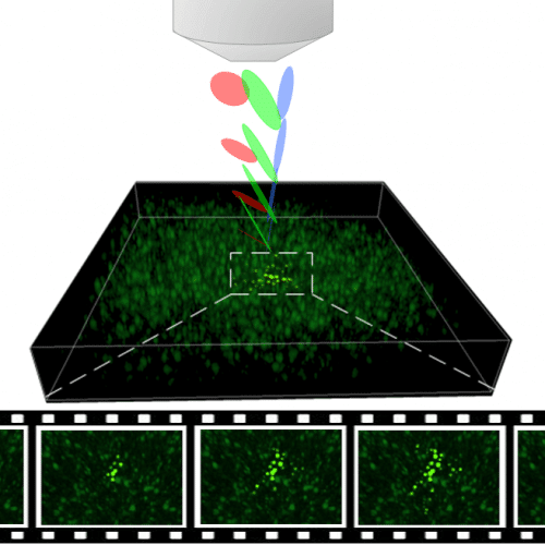
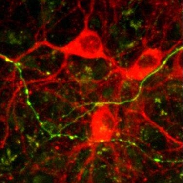
Professor Shai Shem from the Faculty of Biomedical Engineering at the Technion, and the research team that included Dr. Hod Dana, Dr. Anat Marom, Shir Ploch, Roman Dvorkin, and Dr. Inbar Brosh, explain that they grew the advanced cultures in a transparent gel that provides support for the cells and allows for them to connect and create neural networks. "By controlling the conditions of cell growth, we reached a density and composition of cells similar to that found in the human brain, and we also showed the creation and development of links between the cells and the receipt of networks that maintain neural activity," notes Dr. Marom. In order to enable the study of network activity in three dimensions, optical tools are used: the nerve cells in these cultures have been genetically modified so that the activity of the network can be seen under a fluorescent microscope, hence the nickname "optonet".
To observe the activity of the cells in XNUMXD, the researchers developed an advanced imaging system that applies the "temporal focus" technique - a principle from the world of nonlinear optics that was developed about a decade ago at the Weizmann Institute. "The imaging system we developed is a significant step in improving the research ability to observe the activity of multiple brain cells in space and time and thus draw conclusions about brain activity," explains Dr. Dana.
"Through complementary developments in microscopy and neural tissue engineering, we have shown an unprecedented ability to observe more than a thousand cells developing in neural networks with complex self-activity patterns. These innovations open a window for further developments in the field of neural interfaces and additional applications for engineered three-dimensional networks, in the range from basic brain research to examination of the effect of neurological drugs on the activity of nerve cells" concludes Prof. Shaham.
Video: A video describing the culture activity we developed. In the film, you see part of the culture with about 1000 cells, then focus on a small area with synchronized activity of a group of cells (this is one of the unique characteristics of the activity we found in this study). The change in the color of the cells marks their activity which was measured by us.
