The researchers at the Faculty of Biology at the Technion discovered a new geometric structure: the tilted helix
A research group from the Faculty of Biology at the Technion deciphered the action of the protein dynamin in the process that allows the cell to ingest food. The article was published in the journal of the American Academy of Sciences - PNAS.
Biological cells, just like the whole organism, cannot live without eating. Since they do not have an eating organ, during evolution the cells developed a unique and clever technique for ingesting food: endocytosis. In the process of endocytosis, a depression appears in the cell membrane that develops and protrudes inward. At the end of the process, an internal bubble is formed from the membrane, inside which the nutrients are trapped. To complete the act of swallowing, it is necessary to cut the neck of the bubble and release the nutrients into designated organelles in the cell. A key factor in the aforementioned shearing action is a protein called dynamin. The dynamin molecules tighten around the neck of the vesicle and cut it off. This is how the food is released inside the cell.
The problem is that this internal process is very difficult to investigate, and although many research groups have dealt with it, the mechanics of the process have not been clear until now. Hence the importance of the achievement of the researchers of the Faculty of Biology at the Technion, Associate Professor Tom Shemesh and the post-doctoral students Dr. Avihai Kadosh and Dr. Ben Yelin. The model formulated by the three was verified with an atomic force microscope (AFM) by researchers at the University of Geneva.
Associate Professor Tom Shemesh is a physicist by training, so he deals with the world of biology from an unconventional angle, focusing on the physics and mechanics of biological processes. The postdoctoral student Dr. Avihai Kadosh, who completed his master's degree in the Faculty of Physics at the Technion, also took a similar path from physics to biology. "In the dynamics of the cell, there are many aspects of information processing, and they are of course important and critical," explains Prof. Mishna Shemesh, "but we focus specifically on the nominal and formal aspects. In fact we are looking at 'blue collar work' - pushing, pulling and positioning of proteins and other molecules. This is what we did with regard to the helical structure of the dynamin chains in the aforementioned process."
A helix is a recurring motif in the structure of living cells, starting with the double helix of the DNA molecule - the dramatic discovery of Crick, Watson and Maurice Wilkins in 1953 - and ending with many proteins and the cell skeleton. Therefore, say the researchers, it is possible to conclude that this is a structure arising from basic principles common to many systems.
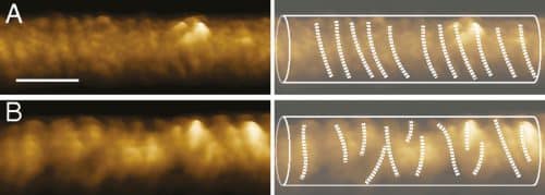
The dynamin chains involved in the endocytosis process are also helical, and the Technion research group's discovery is related to the tilt of the helix at its point of contact with the cell membrane. "In fact, we developed a physical model that links the shape of the protein chain with the mechanical forces that develop in the structure. This model showed that the shape of the dynamin chain and its stability are largely determined by the angle of attachment of the ends of the dynamin protein in the cell membrane. Furthermore, using the model we were able to successfully predict the arrangement of the protein structure in the cells."
Following the findings, the researchers present a new geometric object: the tilted helix. According to Prof. Mishna Shemesh, "if in the past models of intracellular structures were formulated based on classical geometric shapes, here the opposite process happened - biology gave us inspiration for geometry, and this geometry is neither classical nor predictable."
The researchers discovered that the angle at which the dynamin protein is embedded in the membrane is critical to the success of the task - the task of cutting the neck of the membrane and disassembling the dynamin chain to release the food inside the cell. As mentioned, the mathematical conclusion was verified experimentally with an atomic force microscope. Although the research focused on the dynamin molecules, the researchers estimate that the considerations that led to the tilted helix model are universal, and their understanding will lead to the explanation of many phenomena in the cell.
