Back in the XNUMXs, researchers noticed a special pattern of nerve activity in rats - tens of thousands of nerve cells bursting together powerfully in the area of the brain known as the hippocampus. Now the theory has been proven in humans

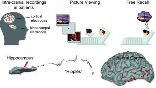
An explorer from another planet who happened upon a football stadium would surely have been surprised to see crowds of people suddenly standing up as one, clapping and roaring with enthusiasm. If he had managed to talk to the fans in their language, they would have told him that they were happy for a goal scored. Similar to the surge of fans, researchers in the 90s noticed a special pattern of nerve activity in rats - tens of thousands of nerve cells bursting together powerfully in the area of the brain known as the hippocampus. However, due to inherent language limitations, the researchers were unable to dub the animals to understand what was going on in their minds at the time of the event. Recently, Weizmann Institute of Science scientists were able to measure the same neural outbursts, known as hippocampal ripples, in humans, and revealed their importance as a neural mechanism involved in the creation and retrieval of memories. These findings are published today in the scientific journal Science.
"This is an amazing event in its strength and speed. An orchestrated burst of about 15% of the hippocampus cells that 'fire' at once and together within about a tenth of a second; Really a show of nervous D-Nor fireworks. Such a dramatic event is not known from other parts of the brain," explains Prof. Rafi Malach from the Department of Neurobiology. Since these bursts were discovered in rats, they have attracted much research interest, and it has been revealed that they characterize sleep and rest states, and play a central role in rodents' spatial memory and navigational abilities. Only recently was it discovered that this group electrical activity also occurs in the hippocampus of monkeys and humans in awake states, but until now it was not known in which cognitive functions it is involved.

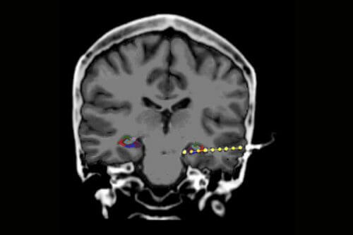
Unlike rodents, the great advantage of brain research in humans is the ability to communicate and understand what is going on in their minds at any given moment. The disadvantage, however, is that in order to record the brain activity in the hippocampus of humans, an invasive intracranial measurement is required. Research student Itzik Norman, from Prof. Malach's group, who led the current study in collaboration with Prof. Ashesh Mehta and his group from the Feinstein Institute for Medical Research in the United States, overcame the obstacle by recruiting subjects who were already undergoing such an invasive examination for medical reasons. These are epileptic patients who do not respond to drug treatment, so electrodes are implanted in their brains in different locations in order to locate the epileptic focus and surgically disconnect it. These patients volunteer of their own free will to participate in brain experiments, while they wait in the hospital for the epileptic seizure to occur.
During the experiment, the subjects saw colorful and detailed images, and were asked to remember them as accurately as possible. Some of the photos presented portraits of famous personalities (from Barack Obama to Uma Thurman), and some presented landscape photographs of familiar sites (from the Statue of Liberty to the Leaning Tower of Pisa). Later, after a short distraction task and while their eyes were covered, the subjects were asked to recall the pictures they saw and describe them in detail. Throughout the experiment, both the subjects' verbal reports and their brain activity were simultaneously recorded. The brain activity was recorded using an electrode placed in the hippocampus and additional electrodes placed in the cerebral cortex.
The cross-check between the brain activity measured by the electrodes and the subjects' reports brought up a series of fascinating findings. First, it was discovered that bursts play an essential role in recall - about a second or two before a subject began to describe a new image, a significant increase in their rate was recorded. It was also discovered that the bursts in the hippocampus reproduce the content of the pictures during recall - pictures that produced more bursts when presented, correspondingly produced more bursts when recalling them. Examining the brain activity measured in the cerebral cortex, showed that the bursts in the hippocampus are synchronized with the brain activity in the visual areas of the cerebral cortex, where the details of the visual memory are apparently stored. Moreover, it is known that the visual areas "specialize" in visual categories - for example, there is a separation in them between representations of people and representations of places. Accordingly, when the subjects were reminded of Barack Obama, or alternatively the Leaning Tower of Pisa, their outbursts were correlated with an increase in neural activity in the relevant area of the cerebral cortex. Norman explains: "An orchestra of several nerve cell populations working in coordination at the moment of recollection, with the hippocampus playing the leading role, was revealed here."

The discoveries greatly expand the existing understanding regarding the role of the hippocampus and how it works. They emphasize the importance of group neural activity, in addition to the unique properties of each individual neuron previously emphasized. "There is a breakthrough here in the understanding of memory mechanisms in the human brain," concludes Prof. Malach. "Creating memories, saving them and retrieving them depend on many other processes, but due to the 'brain drama' of the synchronized bursts, one can definitely estimate that this is a key mechanism in the process of remembering."
More of the topic in Hayadan:
