Dr. Michal Rosen Zvi, director of medical information research at IBM's research laboratories, which is responsible for the field of health in all the company's laboratories around the world, says that the ability developed for Watson will be able to differentiate between photographs of healthy organs and patients, as was done in Haifa in the field of breast cancer detection

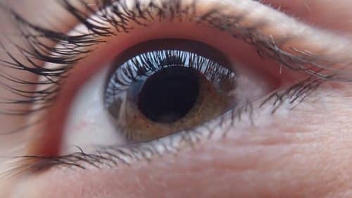
IBM researchers were able to guide the company's cognitive computing system, Watson, to identify retinal anomalies.
The people of the IBM research laboratory in Melbourne, Australia guided the cognitive computer to find abnormal patterns in the images of the retina. These new capabilities will make it possible to offer doctors more comprehensive insights, and speed up the tasks of early detection of patients who are at risk of developing an eye disease such as glaucoma - which is currently the most common cause of blindness in the elderly in the developed world.
Dr. Michal Rosen Zvi, director of medical information research at IBM's research laboratories worldwide, says that "analyzing images requires an ability that has only recently become possible - to distinguish between images that are similar to each other but contain a different feature that was difficult to distinguish until now. A chihuahua dog with black spots on it and a chocolate chip cookie with black spots - can look similar, because once you perceive a two-dimensional image from a certain angle, it is difficult to distinguish between the images."
Distinguish between a normal condition and a cancerous growth
According to her, "In the last three or four years there has been a leap forward in the capabilities of computational learning, learning from examples - a method called deep learning. This method produces a neural network that somewhat simulates how our brain looks at images, and with the help of these neural sequences it is trained to learn to distinguish between types of images. We took this ability in the world of medical images and used it to separate a healthy eye from an eye with glaucoma, and in breast cancer to differentiate between a normal condition and a cancerous tumor."
"With the help of the deep learning method, we were able to achieve higher accuracy between a breast that has a cancerous tumor and a benign one, and between an eye that has diabetes complications called glaucoma and a healthy eye."
The research began in 2015, focusing on optimizing the manual processes currently performed by ophthalmologists. These processes include distinguishing between right-eye and left-eye images, evaluating the quality of the retinal scan, and grading possible indicators of glaucoma.
Glaucoma has been called the "silent thief of sight". Many patients are not diagnosed until the moment when visual impairment is discovered. Glaucoma can be treated - but early detection is a critical matter: today, doctors rely on standard retinal scanning software, which requires an individual examination and analysis by the doctor.
What other actions can be done with this technology?
"The team in Haifa works together with the team in Australia. The application is different - the method is similar, and we share the technology and together develop the capabilities in deep learning", said Rosen Zvi.
"We are now working with Maccabi to integrate the images in which a suspicious finding of breast cancer is found, and we also integrate the information with the patient's medical file and history, as well as follow-up after the image is deciphered, for example a biopsy that showed it was a benign tumor. This requires both deep learning to distinguish between images and learning from their contexts."
Rosen Zvi also said that "we are working on a system that learns from the data of AIDS patients and recommends the cocktail to doctors based on the genome of the virus - the beginning of personalized medicine. Another project is Big Data analysis of epilepsy patients to avoid the need for trial and error until the appropriate treatment is found." Another important announcement from the last few months, she said, "is about cooperation with Teva regarding new uses for old drugs."
95% accuracy rate
According to her, "the researchers used deep learning techniques and technology for the analytical analysis of images, to which they entered 88 photographs of retinas - without identifying details of the people from whom these photographs were taken."
"The research work pointed to Watson's ability to accurately measure the ratio between the size of the optic disc - the blind spot in the retina, where the optic nerve and blood vessels enter the retina - and between the size of the curvature of this area, the rate of which increases with the progression of glaucoma. The ratio between these two values is a main sign of the disease - and the Watson system offers an accuracy rate of 95% in its assessment."
"In addition, the system is able to distinguish between photographs of the right eye and photographs of the left eye, with a level of certainty of up to 94% - and in a way that optimizes and speeds up the work and treatment processes."
In conclusion, Rosen Zvi said that "the research is expected to continue, and to improve Watson's diagnostic capabilities. In the future, it is predicted that this technology will make it possible to diagnose additional eye diseases such as damage to the retina resulting from diabetes, and age-related retinal degeneration (AMD).
On the same topic on the science website:

2 תגובות
Glaucoma is not necessarily a complication of diabetes.