The question: How can the protein structure be studied inside a living cell? The findings: The institute's scientists developed a method for studying the structure of proteins under almost natural conditions, and even managed to apply it to a common protein in frozen human cells. After thawing, the cells returned to full function.

Real life is always more complex than any simulation. The same is true for the structure of proteins, which often changes due to chemical changes, due to contact with other molecules, or as a result of the density and acidity of the fluids inside the cell. Since these structural changes affect the function of the protein, and in order to be able to deeply understand many biological processes, the need arises to study the structure of the protein in its natural environment - that is, inside the cell.
Until now, the existing methods have made it possible to study the structure of proteins only under artificial conditions; That is, after the protein was taken out of the cell and put into the solution, or when it was crystallized for the purpose of continuing the experiments. Today, attempts are being made to determine the structure of proteins inside the cell using methods such as nuclear magnetic resonance. But this technology is not suitable, for example, for the study of large proteins and the study of the structure of proteins in their normal concentration in the cell. Increasing the protein concentration artificially, obviously, creates artificial conditions, and is therefore not an adequate solution.
Now the scientists of the Weizmann Institute of Science have developed a method for studying the structure of proteins under almost natural conditions. As recently reported in the scientific journal Journal of the American Chemical Societyy, Prof. Daniela Goldfarb from the Department of Chemical Physics - in collaboration with Dr. Andrea Martorana and Dr. Akiva Paintoch from her group, Dr. Giuliano Blapardona from the group of Prof. Michael Albaum from the Department of Materials and Surfaces, and together with scientists in Italy - succeeded in implementing a method This is for studying a certain common protein inside the cell.
The scientists used a type of spectroscopy called Double Electron-Electron Resonance (DEER), which is widely used to study the structure of proteins in frozen solution outside the cell. The method is based on measuring the distance between two points in the protein, which are marked by the spin marker - which is a molecule with an odd number of electrons, and because of this it responds to a magnetic field combined with microwave radiation. But the application of this method to study the structure of proteins inside the cell has so far been problematic, because a common spin marker called nitroxide, which has an odd number of electrons on the nitrogen atom, acquires an extra electron inside the cell, and therefore becomes invisible to the magnetic field.
Thanks to their rich experience with spectroscopy in general and with the DEER method in particular, Prof. Goldfarb and her group members decided to use another marker: charged atoms of gadolinium, a chemical element that is known to be stable in the human body, and which is used as a contrast material in magnetic resonance imaging in medicine. Unlike nitroxide, gadolinium does not "disappear" in a magnetic field, but it poses another challenge: it only sends a strong signal in a magnetic field that is ten times stronger than the magnetic field used in normal experiments with the DEER technology. Prof. Goldfarb's laboratory specializes in the application of spectroscopy to problems in biology, and in the past they even developed a spectroscopic technology based on strong magnetic fields for use in other studies of proteins. Now the scientists have adapted the same technology to the DEER measurements in the study of protein structure. They used gadolinium as a marker, first labeled the protein outside the cell, and then introduced it into the cell - where the distances between the two markers were measured.
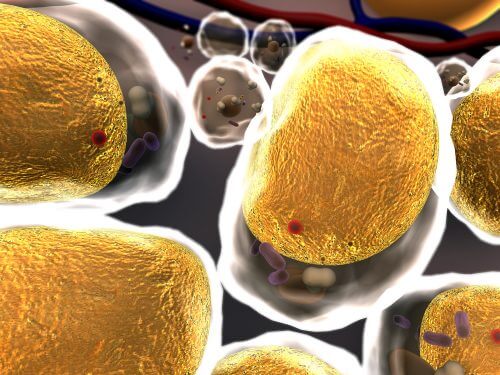
With this method, the scientists were able to study the structure of a common protein, ubiquitin, in frozen human cells, in great detail, down to the scale of ten billionths of a meter. The concentration of ubiquitin inside the cell was similar to that present in its natural environment. But no less important than that: the cells were not damaged as a result of the freezing, and returned to full function after thawing.
Ubiquitin is a particularly important protein - its role in the cell is to mark unwanted proteins destined for degradation - and its structure outside the cell has been known for many years, but the new method will allow biologists to track changes in this structure in a natural environment. Beyond that, it will be possible to apply the method to additional proteins.
The method may advance our understanding of biological processes in which proteins change their structure, including their shape and way of folding, so that at different stages of the process the natural structure may be different from that
studied outside the cell. Structural changes of this type occur in proteins during the transfer of a drug through the cell membrane, absorption of calcium and other substances, and the formation of aggregates in the brain responsible for Alzheimer's disease.

One response
I must not understand something very trivial, but how can a frozen cell serve as a good model for a thawed cell if the structure of the protein "...changes frequently due to chemical changes, due to contact with other molecules, or as a result of the density and acidity of the fluids inside the cell"