More than 100 years after the discovery of the enzymes, most of the studies dealing with their activity are still being conducted in test tubes. Can such experiments really explain what happens in the living cell?

Prof. Gideon Schreiber and his research group in the Department of Biomolecular Sciences at the Weizmann Institute conducted the first experiments monitoring the efficiency of enzymatic reactions in living cells. The findings, Qwere published recently in the scientific journal Journal of Biological Chemistry, show that the catalytic activity occurring in the cell may be chemically identical to that in the test tube - but its efficiency may be less. The explanation for this finding may have extensive implications for both drug design and biochemical research as a whole.
Test-tube experiments are extremely useful, Prof. Schreiber explains, since examining the undisturbed interaction between a clean enzyme and its substrate - the molecules to which the enzyme binds and on which it acts - can shed light on the exact nature of the catalytic reaction mechanism. The enzymatic efficiency in these reactions has been studied in depth, but in the busy, complex and hectic environment of the living cell the rate of these reactions may be different than in vitro conditions.
In order to follow the binding of an enzyme to its substrate and the resulting change in the substrate (the reaction product) in a living cell, the researchers, Dr. Agnes Zutter and Felix Bauerle from Prof. Schreiber's research group, developed methods that would allow them to quantify the process. First, they introduced bacterial genes into a culture of human cells in a petri dish. These genes were engineered to produce enzymes that glow red under the microscope, and the researchers developed an algorithm to quantify the enzymes based on the intensity of the light. The substrate they used glowed bright green in its original state - and bright blue when it became a product; This substrate was injected into each cell individually using a technique known as microinjection.
Later, the researchers produced videos for the cells, which monitored in real time the amounts of the enzyme and the product. Based on the videos, they developed algorithms to plot the reaction rate of these steps, which they compared to more conventional methods.
"The amount of enzyme is almost irrelevant; You can add more 'machines' and get the same production rate. The substrate is the one that limits the efficiency of the process"

The researchers noticed that the amount of the enzyme produced varies from cell to cell. The surprising finding was that the efficiency of the reaction decreased as the amount of enzyme increased. Efficiency, explains Schreiber, is similar to that of a factory: it is based on the total amount of the final product produced in each unit of time, divided by the number of machines. The simulations indicated that the strange result was due to a lack of substrate, but the measurements proved that there was an abundance of substrate in the cells, so the researchers realized that this was not the reason for the finding.
Further simulations indicated an alternative explanation: a significant part of the substrate is bound in other ways, and is therefore not free to react with the enzyme. This explanation was tested by using focused light to "bleach" a small area of the substrate inside the cell and follow the rate at which the dark spot is replenished with the surrounding molecules. According to predictions, these small molecules would have been expected to quickly diffuse throughout the cell, but instead, they moved relatively slowly.
The researchers wondered what was inhibiting the substrate. One of the ways to understand the biochemistry of proteins in the cell is to check where they are located on the continuum between "water lovers" (hydrophilic) and "water haters" (hydrophobic). It turned out that the substrate is close to the hydrophobic end of the scale. This feature makes it somewhat sticky - that is, it tends to bind in a weak bond to several other proteins in the cell and thus becomes "invisible" to the target enzyme, until it is released again.
Prof. Schreiber and the research group compared the hydrophobicity of the substrate to that of drug molecules in common use today, and found that the substrate is within the acceptable range for medical use. "Small molecules that enter the cell, in order to inhibit an enzyme for example, need to be hydrophobic enough so that they can pass through the fatty and hydrophobic outer membrane of the cell, but not so hydrophobic that they get stuck in it," says Prof. Schreiber. "But until now, we have not understood what this property means once the molecule is inside the cell.
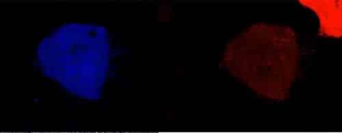
The enzymes (in red) bind to the substrate which is fully converted to the reaction product (in blue) within 1-3 minutes
"One would think that the addition of enzymes - the 'machines' in the 'factory' - would increase output, but the model we developed shows that the amount of enzyme is almost irrelevant; You can add more 'machines' and get the same production rate. It is the substrate that limits the efficiency of the process, but the amount of free substrate may be significantly lower than the amount of substrate actually found in the cell," says Prof. Schreiber.
And there may also be other limiting factors: a drug, for example, needs to penetrate into the cell from the external environment, and may be limited by "gates" in the membrane. In addition, many processes in the cell involve the activity of different enzymes, so that the product of one reaction becomes the substrate for the next reaction. "We still don't know how the efficiency changes at each stage", he adds.
This is the first time that the reaction rates of an enzyme have been tested in living cells. The surprising results of the study may have implications not only for biomedical research, but also more broadly for biochemical research, based on in vitro experiments.
