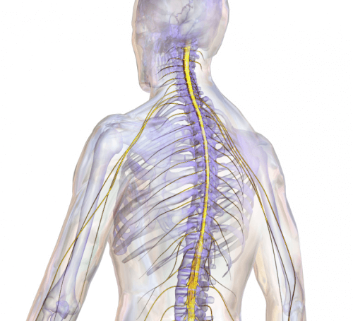
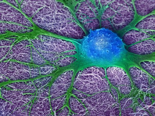
Through a combination of several technologies, the researchers were able to restore a spinal cord with an impressive success rate

A spinal cord injury is aיA severe malignancy that often leads to irreversible paralysis in critical areas of the body. Despite the significant developments in the world of rehabilitation and developments such as nerve transplantation, cellular healing and transplantation of engineered tissues, so far a severed spinal cord has not been fully restored.
Now, collaboration between experts from the world of biomedical engineering and stem cells from Tel Aviv University and the Technion resulted in unprecedented results, when the research group was able to restore the ability to walk to paralyzed rats, through the restoration of a severed spinal cord. The new research raises great hopes, and now follow-up studies are required that will lead to the application of the technology in humans.
The research, recently published in the journal Frontiers in Neuroscience, was led by Prof. Daniel Ofen From the Sackler Faculty of Medicine and the Segol School of Neuroscience at Tel Aviv University, and Prof. Shulamit Levenberg, Dean of the Faculty of Biomedical Engineering at the Technion. The research in the laboratory was led by doctoral students Erez Shur and Javier Gantz.
in a combination of minds
The researchers grew a tissue that would connect the two ends of a severed spinal cord of rats and produce new nerve cells, in order to restore the ability of sensation and motor functions to the rats. For this, a combination of several technologies was necessary.
In the first step, the researchers isolated mature stem cells from the gum area in Prof. Sando Pietro's laboratory from the School of Dentistry at Tel Aviv University. Stem cells from the gum area have a flexible differentiation ability and are relatively easy to produce.
In the second stage, the stem cells were placed on top of the dedicated XNUMXD scaffolds and implanted in the spinal cord, according to the tissue engineering method developed by Prof. Shulamit Levenberg of the Technion. The scaffolding, made of biodegradable materials (bio-compatible) approved for medical use, constitute a three-dimensional environment where the cells can adhere, grow and divide while maintaining proper differentiation and required mechanical properties.
In the third stage, a development by Prof. Daniel Ofen from Tel Aviv University was used, which enabled differentiation of the stem cells into cells that secrete proteins that accelerate the restoration of the nerve cells.

Gradual reconstruction of the spinal cord
The results, as mentioned, are extremely impressive: the transplantation of a piece of 42D engineered tissue restored to 0% of the rats, within about three weeks, the ability to walk, coordination and other motor abilities. This is compared to XNUMX% of the untreated rats from the control group.
According to the standard index of functional rehabilitation (ranging from 21-0), the average score obtained among the treated rats was 9.7. 42% of them achieved a score exceeding 17, which means the restoration of neural control in a way that allows the organism to walk in a coordinated manner, correctly place the foot, the height of the foot above the ground, support weight, general stability and tail-paw coordination. In most of the treated rats, an improvement was also recorded in the sensory responses to external stimuli, following the restoration of the electrical signal pathways between the nerve cells.
Monitoring the rehabilitation process revealed that the transplantation led to the gradual reconstruction of the severed spinal cord, while re-growing axons and nerve cells and inhibiting the formation of a scar. The scaffold is essential to this process, because it directs the growth direction of the nerve cells and axons, and gives the tissue the right balance between flexibility and stiffness. This scaffold gradually degrades and finally disappears about 60 days after transplantation.
The conclusions verify the research hypothesis according to which a specific combination of dedicated scaffolds, stem cells derived from human gum tissue and appropriate growth factors will lead to the regeneration (restoration) of the spinal cord where it has been severed.
The research was conducted with the support of a foundation J&J Shervington, the Israeli Association for Spinal Cord Injury and the National Science Foundation.

4 תגובות
Peace
I wanted to ask, today 4 years after the publication of this article, where do things stand today? Have they done/started experiments on humans?
Very impressive and encouraging, the sky is the limit.
Stunning!
For the attention of the disabled! Don't despair! Do not lose hope!
A few years ago it was also reported in the media about a paralyzed man who, thanks to the treatment and transplantation of nerve cells taken from his nose (our nose is extremely rich in nerve cells), was able to get up and walk with a walker.
Medicine is advancing in this field and with God's help I am sure that the day will come when we will see paralyzed people who have returned to full function.
As far as I know they managed to cure cancer, diabetes and do amazing things with mice.
with less humans….