This is according to a study carried out at the Weizmann Institute. Future application of this method will allow an unprecedented glimpse into what is happening in the brain and other organs
Many substances in our body are in too low a concentration to be observed in magnetic resonance imaging (MRI). Weizmann Institute of Science scientists and their research partners have shown that some of these substances can be "observed" indirectly through MRI monitoring of their interactions with the water molecules surrounding them. The application of this method in extremely strong magnetic fields may allow an unprecedented glimpse into the metabolism in the brain and other organs, thus possibly allowing early detection of cancer, degenerative diseases of the nervous system and other diseases.
MRI makes it possible to simulate living tissue by placing it in a static magnetic field and measuring the oscillation frequency of the atoms in the tissue - usually hydrogen atoms. In order to increase the sensitivity of the MRI and enable the detection of low concentrations of essential chemical compounds known as metabolites, scientists tried to use, among other things, a method called CEST (chemical exchange saturation transfer), which relies on tracking loose hydrogen atoms that are able to break the their connections with their original molecule and create connections with the water molecules around them. Such exchanges occur continuously in the body at a rate of several thousand per second. Since our body contains much more water than metabolites, tracking the new bonds that are created as a result of these exchanges may produce magnetic resonance "signatures" that are much stronger than in a normal MRI test, thus indicating the existence of low concentration metabolites.
In an era when more powerful magnets will be available for use in humans, we may be able to use CEST for the early detection of brain tumors."
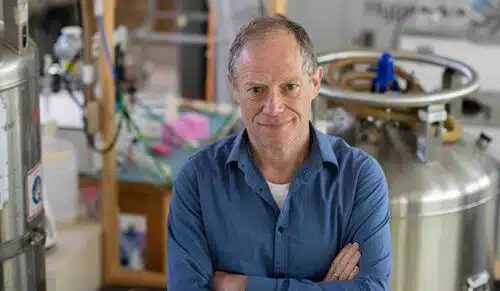
In the past, when scientists tried to use CEST in 3 Tesla MRI systems, which are common in hospitals and clinics, the effectiveness of the method was limited. In the new study, which was recently published in the scientific journal NMR in Biomedicine and appeared on the cover of the journal, the scientists showed that the method works much better in stronger magnetic fields. To examine strong magnetic fields, Prof. Lucio Friedman and postdoctoral researcher Tanji Russell from the Department of Chemical and Biological Physics of the Samuel Grant Institute teamed up with Dr. Jens Rosenberg from the National Laboratory for Strong Magnetic Fields in Tallahassee, Florida, USA, where The strongest MRI magnet in the world operates - with a power of 21 Tesla. With the help of this magnet, which is currently intended for animal research only, the scientists discovered that every increase in the strength of the magnetic field translated into a squared increase in the "signatures" obtained through CEST; For example, doubling the field quadrupled the signatures.
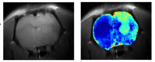
The scientists showed that at these intensities, the method effectively reveals metabolic changes in the brain of a rat, which indicate a cancerous tumor of the glioblastoma type. The method also made it possible to distinguish damage to the integrity of the brain tissue following the tumor; The glioblastoma does not immediately destroy the tissue, but it makes it more "watery" than usual. Changes of this type are almost invisible on conventional MRI, but were clearly observed with CEST.
In an era when more powerful magnets will be available for use in humans, we may be able to use CEST for the early detection of brain tumors. In follow-up studies, the scientists plan to use two powerful magnets that were recently installed at the institute - an MRI device with a power of 7 Tesla approved for use in humans, and a power of 15.2 Tesla for animal research - to study other types of cancer and enable the early detection of cancer in stages where it cannot be detected in the future in a normal MRI. CEST may also be used to detect and diagnose other diseases, including neurodegenerative diseases of the nervous system.
The magnetic field where the research was carried out is 600,000 times stronger than the Earth's magnetic field, which has a strength of 0.000035 Tesla.

2 תגובות
Can tests with very high magnetic fields cause damage, for example I read that it can lead to epileptic seizures? Will it be possible to prevent it?
Very interesting. Will it be possible to understand the mechanism of diseases such as multiple sclerosis, depression, Parkinson's, etc.? And can this help us in the future to effectively treat these diseases?