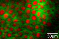
The method, based on stimulated Raman scattering (SRS), works by measuring the vibrations of the chemical bonds between the various atoms.

A new method of highly sensitive microscopy, developed at Harvard University, could greatly expand the boundaries of current biomedical imaging, allowing scientists to pinpoint the exact location of drugs and their breakdown products in living cells and tissues without the need to use any type of marker. Fluorescent.
The method, based on stimulated Raman scattering (SRS), works by measuring the vibrations of the chemical bonds between the different atoms. This type of microscopy could provide scientists with a new form of three-dimensional and real-time biological imaging without the need for fluorescent markers that could themselves inhibit many biological processes. The research, led by scientist X. Sunney Xie, is described in the scientific journal Science.
"SRS microscopy is a major breakthrough in biomedical imaging and a breakthrough in the real-time study of metabolic processes in living cells," says principal investigator Xie, a professor of chemistry and biochemistry in the Faculty of Arts and Sciences at Harvard University. "We have already used this method in the past to map lipids in a living cell and to measure the release of drugs into the interior of living tissues. There are only two earlier examples of the influence of this microscopy in the fields of medicine and cell biology."
The scientists' research, mapping unsaturated and saturated fats in living cells, provides new tools for further metabolic studies of omega-3 fatty acids, which are required by the human body for its normal activity, but are not produced in it. [From Wikipedia: "A suitable dose of omega-3 in the diet is associated with positive health effects, and is used as a common dietary supplement. However, it should be remembered that its effects are still under research, and there may be future studies that will contradict these connections. Its positive effects include improving the circulatory system and reducing the risk of coronary heart disease, reducing the risk of cancer, an antidepressant effect and improving attention span. At the same time, a number of negative effects were also found such as: increased bleeding, especially at a dose over 3 grams per day, cerebral bleeding and reduced glycemic control in diabetics."].
Despite the increase in the number of solid evidence indicating that acids of this type provide many health benefits such as suppressing inflammation, reducing blood triglyceride levels and helping to eliminate cancer cells, almost nothing is known about the breakdown and processing of these types of fatty acids in our body.
"Our diet has undergone significant changes in recent decades," notes the lead researcher. "As a unique method capable of distinguishing the distribution of lipids in living cells - and capable of differentiating between different types of lipids - SRS microscopy could prove to be extremely useful in helping to understand and treat the growing imbalance between saturated and unsaturated fats in our diet." The method can help to a considerable extent also in the field of imaging the nervous system since the nerve cells (neurons) are wrapped in a fatty sheath known as myelin.
The researchers' use of this method to examine skin tissues could open new horizons in the field of drug development as well. The researchers used this method to observe the absorption efficiency of retinoic acid, a drug for local use against acne (pimples), into the skin cells. They also used the method to track the deep penetration into the skin of the substance dimethylsulfoxide (DMSO), a compound added to many topical medications and ointments to increase absorption.
Scientists currently use a variety of methods to monitor biocompounds, but most of them have significant limitations that are avoided in SRS spectroscopy. A certain protein from jellyfish that was first discovered in 1962, green fluorescent protein (GFP), is now widely used as a marker to monitor the activity of bio-compounds. This labeling method provides sharp images, but the bulky protein may interfere with delicate biological processes, especially in cases involving smaller biocompounds. In addition, the unique fluorescence of this protein fades over time and as a result does not allow long-term follow-up.
Similar to the SRS microscope, infrared (IR) microscopy and Raman microscopy measure the vibration of the atomic bonds between the different atoms. However, they are very sensitive imaging methods because they require dry samples or high laser power, requirements that limit their use in animal samples. Another microscopy method developed in this research group - "Coherent anti-Stokes Raman scattering" (CARS) does not provide sharp enough images for most compounds.
