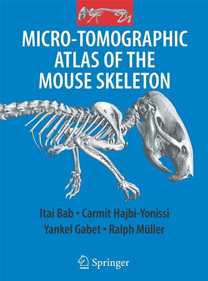
"Micro-tomographic atlas of the mouse skeleton", the result of the work of Hebrew University researchers, offers an accurate and detailed mapping of the mouse skeleton

"A micro-tomographic atlas of the mouse skeleton" is a new book that reviews the structure of the mouse skeleton in a detailed and focused way, using the computerized micro-tomography technology, which allows the presentation of details down to the size of thousandths of a millimeter. The book, which is published by the prestigious scientific publishing house Springer, New York, is the result of the joint work of a team of researchers from the bone laboratory at the Hebrew University: the head of the laboratory Professor Itai Bab, Dr. Carmit Hajabi-Younisi and Dr. Yankel Gavet. Professor Ralf Müller from the University of Zurich also participated in the writing of the book.
The new atlas indicates a great similarity between the human skeleton and the mouse skeleton, except for the structure of the face and the hands and feet. Like humans, for example, mice suffer from skeletal diseases such as osteoporosis. The meaning of this similarity is that findings and conclusions arising from research on the anatomy of the mouse are highly relevant to research on the human body: findings from research in mice on topics such as development, drug testing and skeletal diseases have far-reaching implications for the corresponding topics in humans.
According to Professor Babb, in the last century several anatomy books describing the skeleton of the mouse were published, which contained drawings based on visual observations only. Today, these books are not suitable for modern research: they contain inaccurate information, not detailed enough, and the various parts of the skeleton are only described in an external description.
Professor Babb explains that in the last decade the computerized micro-tomography method was developed, which became the leading technology for imaging the skeleton. With this technology, two-dimensional and three-dimensional imaging is obtained showing details up to the size of six thousandths of a millimeter. The new atlas presents approximately 200 XNUMXD and XNUMXD images of the external outline of all parts of the mouse skeleton, and images of the internal anatomy of the bones. Also, the three-dimensional relationships between the bones in different positions of the joints are shown. A special chapter, based on skeletal measurements at different ages, describes the development of the skeleton and its aging, as well as the differences between males and females.
The computerized micro-tomography system used by the researchers was purchased by the Hebrew University with the help of a grant from the National Science Foundation.

6 תגובות
Certainly mice have bones, they are vertebrates and even mammals.
I haven't been answered yet
The question is do mice have bones
I didn't understand anything they said
Yo, what poor mice! Now they will do more disgusting experiments on them
Come on berry berry mice
come come ancestor
Shimon Peres evolved from Donald Duck
We all evolved from Mickey Mouse.