"We have a model here that is not exactly a brain, but it is a good model for the development of the brain," says Prof. Orly Reiner, "and we now understand better why the brain in patients is smooth and not folded

In Western philosophy, since the days of Aristotle, the idea that humans come into the world "tabula rasa" (Latin: flat board) - that is, without prior knowledge - has gained traction. However, biologically speaking, the highly folded human brain, that walnut-like structure, is far from a smooth slab. In fact, babies born with a smooth brain suffer from a severe developmental disorder - they cannot talk or walk, and they usually die in childhood. Despite the understanding that the winding surface plays an important role in the brain of humans, so far no explanation has been found regarding the way these folds develop. In a new study, published today in the scientific journal Nature Physics, Weizmann Institute of Science scientists used an innovative method to grow miniature human "brains" in the laboratory, in order to simulate and reveal the physical and biological mechanisms that cause the formation of the cerebral folds.
In the last decade, there was a breakthrough in the study of brain development, when scientists in the laboratories of Prof. Yoshiki Sasai from Japan and Prof. Jürgen Knoblich from Austria for the first time grew human brain-like structures, called organoids, from embryonic stem cells. This scientific development aroused great interest among neuroscientists, and laboratories around the world adopted the method. "We were very enthusiastic about the new method," says Prof. Orly Reiner from the Department of Molecular Genetics, "we hoped that it would allow us to understand developmental processes of the human brain that cannot be understood using model animals such as mice, because in them the normal structure of the brain is smooth." But despite the enthusiasm, Dr. Eyal Kretzbron, then a postdoctoral researcher in Prof. Reiner's laboratory, soon discovered that the new method also had limitations: "We found a huge variation in the size of the organoids: some of them reached the size of several millimeters, and some were much smaller. Moreover, when you cut them open, you find that in the absence of blood vessels and a proper supply of nutrients, the organoids begin to 'die from within'. Another significant obstacle is the thickness of the resulting tissue, which does not allow for optical imaging and real-time microscopic monitoring of the growth processes."
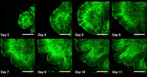
To overcome the limitations, Dr. Kratzbron developed a new approach to grow the organoids. The researchers limited their growth along the height axis, and as a result, the cells organized themselves in the form of a thin, round structure that wraps around a narrow space, which is more reminiscent of a pita. Thanks to the thin tissue, real-time visualization of the growth was possible, and more importantly - it was possible to supply food to all the cells. In the second week of the development of the "brains", Dr. Kretzbron identified folds that got deeper and deeper. "This is the first time folds have been identified in organoids," he says, "and this is probably thanks to the architecture of our system." Dr. Kratzbron, a physicist by training, turned to physical models of the behavior of elastic materials in an attempt to understand how the folds are formed. Physically, folds appear on the surface as a result of mechanical instability when opposing compressive forces are applied to an elastic material; Opposing compression forces can be created, for example, as a result of uneven "swelling". Indeed, the researchers identified the action of two opposing forces in the organoids: on the one hand, the contraction of the cellular skeleton in the core of the organoid, and on the other, the expansion of the cell nuclei near the surface. In other words, the outer part of the "pita" grows faster than the inner parts.
At this point, Prof. Reiner was not convinced that the observed folds actually simulated the brain's developmental process. To test this, the researchers grew organoids from stem cells into which a mutation was introduced that causes smooth brain syndrome. Already in 1993, Prof. Rainer identified the LIS1 gene, whose mutation causes this syndrome one in 30,000 births. This gene is responsible for many essential functions, and is involved, among other things, in the migration of nerve cells, and in the control of the cellular skeleton and intracellular molecular motors. A few days later, the mutant organoids grew to dimensions similar to the normal ones, but only a few folds were formed in them - with a huge difference in the wavelengths. The researchers hypothesized that the difference in folds is due to different physical properties of the cells. Using an atomic force microscope, and with the help of Dr. Sidney Cohen from the Department of Chemical Research Infrastructures at the Weizmann Institute, it was discovered that the modulus of elasticity of the normal cells was approximately twice that of the mutant cells. Or, in other words, the mutant cells were simply softer. Prof. Rainer says: "We discovered a very significant difference in the physical properties, but the biological properties were also different. For example, the speed of movement of the cell nuclei to the nucleus was much slower in the mutant cells, and there was a very significant difference in the proteins of the intercellular tissue."
Even before the research was published, there was interest in the scientific community in the new approach to grow organoids. "We have a model here that is not exactly a brain, but it is a good model for the development of the brain," says Prof. Rainer, "and we now understand better why the brain in patients is smooth and not folded." The researchers plan to continue developing the model, in an attempt to understand other diseases associated with poor brain development, including microcephaly (small brain), epilepsy and schizophrenia. Prof. Yaqub Hana, who specializes in the treatment of embryonic stem cells, and research student Aditya Kashirsgar, from Prof. Rainer's group, also participated in the study.
36 hours in the life of a brain organoid (in red - the cell nuclei, in green: the cytoskeleton). The folds appeared on the surface after uneven "swelling".
5 hours in the life of a "smooth brain" organoid (in red - the cell nuclei, in green: the cytoskeleton). The mutant organoids grew to similar dimensions to the normal ones, but only a few folds formed in them

3 תגובות
folds
I think I understand why there are no multiples (maybe I'm wrong) and it's taken from acceptance in artificial intelligence.
I don't think we have any control over everything that goes on in our minds. For all that it implies!?…