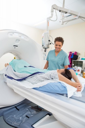
Researchers are reevaluating the risks associated with radiation exposure following medical imaging tests

Ever since doctors began routinely using computed tomography (CT) scans about forty years ago, researchers have been concerned that the medical imaging process could increase subjects' risk of developing cancer. Computed tomography scans bombard the body with X-rays that can damage DNA and lead to the creation of mutations and subsequently to the creation of tumors.
Doctors have always believed that the advantages outweigh the disadvantages. The X-rays that rotate around the head, chest or other organ create a three-dimensional image that is much more detailed than the image obtained from a single X-ray. But one such scan exposes the body to a level of radiation 150 times to 1,100 times higher than that to which the body is exposed in a single X-ray. This level of radiation is equivalent to the level of radiation to which the body is exposed during an entire year from both natural and artificial environmental sources.
Several studies published in the last ten years raise the concerns again. Researchers from the American Cancer Institute estimate that it will be possible to attribute 29,000 of the future cancer cases in the US to more than 72 million computed tomography scans performed in the country in 2007. This is therefore about 2% of all 1.7 million cancer cases diagnosed in the US each year. Another 2009 study of San Francisco Bay Area medical facilities also found an increased risk of cancer: one additional case of cancer for every 400 to 2,000 routinely performed chest CT scans.
The reliability of these estimates depends, of course, on how scientists initially measure the relationship between the level of exposure to radiation and the risk of getting cancer. In fact, most of these estimates are based on a potentially misleading database: the number of cancer cases among survivors of World War II atomic bombings.
"It is very problematic to deduce from the data on the survivors of the atomic bombings the true risk of getting cancer from a computed tomography scan," says David Richardson, an associate professor of epidemiology at the Gillings School of Public Health at the University of North Carolina who has conducted research on the survivors of the nuclear bombs.
About 25,000 survivors of the bombings were exposed to low levels of radiation equivalent in value to the level of one to three computed tomography scans. However, the number of cancer cases that developed among this group is not high enough to provide the statistical significance needed to conclude from these data about the increase in the risk of getting cancer among the entire population today. In view of this problem, as well as due to the renewed concerns and the lack of a mandatory standard for performing safe CT scans (as opposed to other medical imaging operations such as mammography), many research groups around the world have begun to reassess the risk associated with computed tomography tests based on more reliable data.
A growing number of doctors and medical associations are not waiting for decisive conclusions about the health risks associated with computed tomography and have already started looking for ways to reduce radiation levels. For example, two radiologists from Massachusetts General Hospital claim to be able to reduce the X-ray dose of at least one type of scan by 75% without significantly compromising the quality of the resulting image. Similarly, some medical societies are trying to reduce the number of unnecessary scans and get doctors to reduce the level of radiation they use during scans.
Outdated information
For obvious ethical reasons researchers cannot simply expose people to radiation just to test for increased cancer risk. Because of this, scientists used data from survivors of the atomic bombs dropped on Hiroshima and Nagasaki in August 1945. Between 150,000 and 200,000 people were killed directly during the bombings and in the months that followed. Most of the people who stayed up to a kilometer away from the explosions died from severe radiation poisoning, skin loss or incinerators that erupted immediately after them. Some people who were less than 2.5 kilometers away from the bombing site lived for years after being exposed to different levels of gamma radiation: from a very high level of more than 3 Sievert (Sv) which causes burns and hair loss, to a low level of 5 thousand Sievert, which is Corresponding to that of the average exposure in a CT scan accepted today (between 2 and 10 thousand Sieverts). Sievert is an international unit for measuring the effect of different types of radiation on tissues. One sievert of gamma radiation causes tissue damage equal to that caused by one sievert of x-ray radiation.
A few years after the bombing, scientists began collecting data on the rates of disease and death among the more than 120,000 survivors. The results showed for the first time that the increase in the risk of getting cancer depends on the level of radiation to which the body is exposed, and that even exposure to a very low level of radiation increases the risk. Based on these data, a report by the US National Research Council in 2006 estimated that exposure to a radiation level of 10 thousand Sieverts, which is the level of a computed tomography scan of the abdomen, increases the risk of developing cancer during life by 0.1%. Using the same database, the US Food and Drug Administration estimated that exposure to a radiation level of 10 thousand Sieverts increases the risk of developing fatal cancer by 0.05%. Because these rates are tiny compared to the number of cancers in the general population, they don't seem alarming. Every person in the US has a 20% chance of dying from cancer. Therefore, one CT scan can increase the average chance of developing a fatal cancerous tumor from 20% to 20.05%.
But all these estimates suffer from a common serious flaw. The number of cancer cases among the survivors who were exposed to a radiation level of less than a tenth of a Sievert, a level of radiation that is equivalent to or slightly greater than that caused by a single computed tomography scan, is so low that it is impossible to determine whether there has been an increase in the risk of developing cancer compared to the general population. To compensate for this lack of statistical significance, entities such as the US National Research Council based their estimates on the survivors who were exposed to radiation levels between 0.1-2 Sievert. The fundamental assumption on which these estimates are based is that the relationship between the level of radiation to which they are exposed and the risk of getting cancer remains constant at high radiation levels and at low radiation levels. But this assumption is not necessarily correct.
Another factor that complicates the picture is the fact that the nuclear bombs exposed the entire body to one burst of gamma radiation, and in contrast, patients are exposed to several computed tomography scans that focus X-rays on a specific area of the body. At the same time, the survivors of the nuclear bombs lived on a less good diet, and did not have good access to medical services compared to the population in the US today. Therefore, it is possible that exposure to the same level of radiation will cause a sharper increase in disease rates among the survivors of the bombs than among US citizens today.
Reduce radiation levels
In order to reliably assess the risks of exposure to low levels of radiation and to establish acceptable safety rules regarding computed tomography, researchers stopped relying on the data obtained from the survivors of the nuclear bombs, and began to directly examine the number of cancer cases among people who underwent computed tomography scans. Several such studies that looked at the incidence of different types of cancer among those who underwent screening will be published in the coming years.
In the meantime, other researchers began to test whether it was possible to obtain better images using radiation levels lower than those used in a typical test. Sarabjit Singh, a radiologist at Massachusetts General Hospital, and his fellow radiologist Manudeep Kalra, found an original way to conduct such research. Instead of irradiating live volunteers, they perform CT scans of corpses. In this way they are not limited in the number of scans. They can also perform postmortems to check if the scans they performed accurately located medical problems in the body.
So far, the two researchers have discovered that they can diagnose abnormal tumors in the lungs and perform routine chest examinations using radiation levels 75% lower than those currently accepted. Following this, Massachusetts General Hospital adopted their testing protocol. Singh and Clara are currently disseminating their findings among radiologists and x-ray technicians in hospitals and imaging centers throughout the US and the world.
Medical associations also began to participate in the effort. Since the US Food and Drug Administration does not supervise the use of CT machines and does not set the radiation levels allowed in the tests, different centers use a wide range of radiation levels, some of which are apparently too high. Over the past year, Singh says, the American Association of Medical Physicists has published guidelines for the regulation of computed tomography procedures, which will lead to the restraint of imaging centers that use abnormal levels of radiation. Also, more and more imaging centers across the US are receiving certification from the American Society of Radiology, which sets threshold standards for the permitted radiation levels and evaluates the quality of the images obtained as a result of the imaging process. As of 2012, outpatient clinics in the USA must receive this certification if they wish to receive reimbursement from Medicare, the public medical insurance of the USA.
However, it doesn't matter how much the imaging institutes reduce the level of radiation they use, another problem still remains. Many times unnecessary computed tomography scans are performed and the patients are exposed to unnecessary radiation. Bruce Hillman of the University of Virginia and other researchers are particularly concerned that emergency room doctors are ordering too many such scans because of their need to make decisions under pressure. A survey conducted in 2004 revealed that 91% of emergency room doctors do not believe that computed tomography increases the risk of getting cancer. But the doctors, as well as their patients, are finally starting to digest the message. Analysis of Medicare data from 2012 shows that the upward trend in the number of CT scans has begun to slow down and may even reverse.
"Researchers are still trying to determine if there is a health risk in CT scans," says Daniel Prosh, chief pediatric radiologist at Duke University Health Center. "But the safest way is to assume that there is no safe dose of radiation. And even if in 20 years we discover that a small amount of radiation is not harmful, what will we lose by trying to minimize our exposure to radiation today?"
____________________________________________________________________________________________________________________________________________________________________
on the notebook
Karina Stores (Storrs) is a freelance science and health reporter. Her articles have been published in popular science magazines, The Scientist and Health.com, among others.
The article was published with the permission of Scientific American Israel
