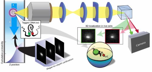
The research group of Dr. Yoav Shechtman from the Faculty of Biomedical Engineering dismantled an existing imaging device worth hundreds of thousands of dollars and reassembled it. The result: a device that performs 1,000D imaging of XNUMX cells per minute

Researchers at the Technion have developed an innovative method for XNUMXD imaging of nanoscale processes - for example, inside living cells in a flowing liquid. The research, which was carried out in the research group of Dr. Yoav Shechtman from the Faculty of Biomedical Engineering led by postdoctoral student Dr. Lucian Weiss, was recently published in the prestigious journal Nature Nanotechnology.
According to Dr. Shechtman, "Our goal is to enable XNUMXD imaging inside living cells under conditions that simulate their natural environment, and just as importantly - to do it at a high output (fast rate). This is a huge challenge, because XNUMXD microscopy often requires a long time and some kind of scanning. Here we did it from a single image and while flowing."
The experiments in the new system were conducted on DNA molecules in live yeast cells and on white blood cells with engineered nanometer particles provided by the laboratory of Prof. Avi Schroeder from the Wolfson Faculty of Chemical Engineering at the Technion. The gist of the achievement: 1,000D imaging, with high resolution, in more than XNUMX cells per minute.
Dr. Shechtman explains that "this success can have very important applications in the context of basic science, for example in understanding the three-dimensional structure of DNA inside a living cell, but also in the field of nanomedicine, that is, in medical treatment based on engineered nanometer particles As developed in Prof. Schroeder's laboratory. Thus, for example, we can use the new technology to measure the rate of absorption of such therapeutic particles in the living cell, track their distribution in the cell and monitor their effects on it. Today there are methods for mapping and measuring cells, but the methods characterized by high throughput provide a partial and two-dimensional image. Our technology combines the advantages of the various methods and provides a XNUMXD image at a high rate."
The innovative technology is based on the re-engineering of ImageStream - an advanced imaging device purchased for hundreds of thousands of dollars by the Lori Lockey Interdisciplinary Center for Life Sciences and Engineering at the Technion. This device combines two different technologies - flow cytometry and fluorescent microscopy - and thus enables the analysis of cells at a high rate.
According to Dr. Shechtman, "The sampling rate and the amount of sampled cells are very important in the biological context, because a biological experiment is always 'noisy' and imprecise, and to draw any quantitative conclusion we need statistics on large amounts. There are cases where, due to a low sampling rate, it is impossible to collect such statistics, because by the time you finish collecting the information - the phenomenon under study has already changed. That's why it's important to have technology that enables sampling at a high rate."
ImageStream is used for many purposes including characterizing populations, diagnosing medical conditions and testing new drugs. "It's an excellent device," says Dr. Shechtman, "but it has a significant limitation - it only allows images of objects in two dimensions, meaning it only gives us a two-dimensional mapping of them. Obviously this is a problem, because objects in the real world are XNUMXD. Even if we only want to check distances inside some particle, a two-dimensional measurement is not enough, because even in the depth dimension there is a certain distance between every two points."
And that was exactly the main technological challenge in this study - to turn ImageStream into a 600D imaging device. "For that we had to open it and reassemble it with our unique optical system. You have to remember that this is a $XNUMX device, so the agreement of the imaging center at the Lockey Institute to do this was not obvious, but from the moment we opened the device and looked inside, it was clear to us what needed to be done (without harming)."
The research group mounted on ImageStream the technology it has developed in recent years - technology for localization microscopy based on wavefront design. It is in fact a controlled deformation of the optical system, which allows to map the positions of particles in the three-dimensional space. This technology is based on photographing dye molecules that are embedded in the sample and mark important locations within it (eg cell nuclei). Based on the shape obtained by the camera, after passing through the distorted optical system, the system analyzes the XNUMXD position of the object being examined.
So far, the technology has been used for XNUMXD imaging of static systems, and the connection to the cytometry device gives it the ability to map cells in flow. This connection, which in itself is a huge technological challenge, led to success in sampling at a very high rate - thousands of cells per minute. The Nature group, as mentioned, expressed confidence in the breakthrough by publishing in its leading journal, and the researchers estimate that the technological achievement will lead to important scientific and applied developments in biological and biotechnological research, medical diagnosis and the development of new medical treatments.
Dr. Onit Ellof, Dr. Sara Goldberg, and doctoral students Yael Shalu Ezra, Boris Fredman and Omar Adir also participated in the work.
More of the topic in Hayadan:

One response
Sorry for the question, but what can be concluded from such blurry pictures? is it sealed Is it a molecule? Is it a UFO? I see an unidentifiable spot in the picture, I don't know what exactly can be done with it.