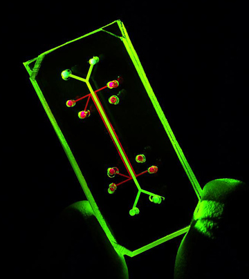
Researchers from Harvard University, in collaboration with researchers from Boston Children's School, have developed a device that mimics a living, breathing human lung on a microchip. The device, the size of a regular pencil eraser, works like a lung in the human body and consists of human lung cells and blood vessels.

"The ability of the "lung-on-a-chip" device to provide a prediction regarding the absorption of airborne nanoparticles and to mimic the inflammatory response that appears after exposure to various pathogens, provides proof of principle for the idea that organs-on-a-chips could replace many of the experiments in the future," says one of the research partners. The research findings were published in the scientific journal Science.
Until today, microsystems of engineered tissues were limited in their activity both mechanically and biologically, explains the researcher. "We are not really able to fully understand how biological systems work unless we integrate them into the physical context of real living cells, tissues and organs."
With each breath of the person, air enters the lungs, fills microscopic pockets called nadia and transfers oxygen through a permeable, flexible and thin membrane found on the surface of the lung cells into the bloodstream. It is this membrane - a three-layered interface of lung cells, permeable extracellular matrix and capillary blood vessels - that does most of the dirty work of the lung. Moreover, this lung/blood interface recognizes invaders such as inhaled bacteria or toxins and activates the immune response.
This lung-on-a-chip microdevice uses an innovative approach to tissue engineering by placing two layers of living tissue—the inner lining of the lung's air sacs and surrounding blood vessels—on top of a flexible, porous substrate. Air is pumped into the lining cells of the lung, rich culture medium is pumped through the capillary channels to mimic blood flow, and a cyclic mechanical stretch system mimics breathing. The device was produced using an innovative manufacturing method that uses transparent flexible materials.
"In this study, we were influenced by the activity of breathing in a human lung, which is carried out by receiving the vacuum that is created when our chest expands, drawing air into the lungs and causing the walls of the lungs to stretch," says one of the researchers. "Our use of vacuum to mimic our microengineered system was based on design principles derived from nature."
In order to determine how well the device mimics the natural response of living lungs to stimuli, the researchers tested the device's response to an inhaled live vocal cord bacteria. They injected the bacteria into the air channels on the side of the device containing the lung cells and at the same time flowed white blood cells through the channels on the side of the device containing blood vessels. The lung cells recognized the bacteria and through the porous membrane activated the white blood cells, which in turn activated the immune response that eventually caused the migration of the white blood cells towards the air chamber and the destruction of the bacteria.
"The ability to reproduce the real mechanical and biological conditions in living systems is a fascinating invention," notes the researcher.
The researchers put a variety of nanoparticles into the air chamber. Some of these particles are present in commercial products; Others are found in air and water pollution. Several types of these nanoparticles penetrate the lung cells and cause them to generate a large amount of free radicals and trigger an inflammatory response. Many of the particles passed through the lung model into the bloodstream, and the researchers found that mechanical breathing significantly increases the absorption of nanoparticles. These new findings were verified in mice.
"This lung-on-a-chip is simple and combines several technologies in an inventive way," explains the researcher. "I believe that the device will be useful in testing the safety of various substances for the lung and I can imagine the growth of other similar applications, such as in the field of examining the mechanism of lung activity."
According to the researchers, it is still too early to predict how successful this field of research will be. And yet, the ability to use human cells to reproduce the mechanical properties and chemical conditions of organs could provide a real revolutionary change in the field of drug discovery, the researchers note.
The researchers have not yet demonstrated the system's ability to mimic the passage of gas between the air voids and the blood circulation - a key function of the lungs, but, the researchers say, they are looking into it right now.
The team of researchers is also working on the development of models for other organs, such as intestines-on-a-chip, as well as bone marrow and even cancer models. In addition, they are examining the potential for integrating separate organ systems.
For example, another research team at the same institute developed a breathing heart-on-a-chip. They hope to combine their lung-on-a-chip with the heart-on-a-chip. The combination of the engineered tissues could be used to test inhaled drugs and to identify new and more effective medical treatments that lack side effects harmful to the heart.

One response
For my general information: what about artificial lungs?
It seems that from a practical point of view the rejection of a transplanted lung is much more problematic than the transplant itself.
The lungs are a passive organ, and the passage of gases is done passively. If so, why is it so difficult to develop a structure with endothelial channels (capillaries) lining selective membranes?