The brain systems involved in the process of transitioning from wakefulness to sleep * Research using EEG neurofeedback and functional imaging made it possible to measure the change in a person's awareness during the process of falling asleep (in order not to alarm any reader, I would change the order: "The change in a person's awareness during the process of falling asleep is measured using neuro EEG technologies feedback and functional imaging")
Dr. Sivan Kinreich
A changing mental state, such as shifting the focus of awareness from the outside world to the inside, is a daily phenomenon that occurs when a person goes to sleep. Difficulty in such a transition between states of consciousness can be reflected in problems with falling asleep, which is typical for people suffering from disorders such as depression, anxiety or healthy people experiencing periods of stress. Difficulties with anesthesia are also common with increasing age. Despite the importance of understanding the brain processes that accompany the change in the state of arousal, there are not many brain studies of the phenomenon that have been done on people. This may be due to the difficulty of controlling the process in the laboratory. For example, most studies require the subjects to avoid sleep for a long time before the experiment in order to bring about a state of sleep deprivation and thus a relatively quick transition to a state of sleep will be possible.
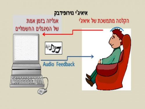
One of the more well-known ways to assess the state of wakefulness during the transition to sleep is by recording EEG electrodes placed on the scalp and analyzing the change in the frequency spectrum. The neural brain activity produces an electrical signal that is picked up by the electrodes and can indicate a change in the mental state. Studies using this technology found that during the transition to sleep, the frequency known as alpha decreases in intensity while the intensity of the theta frequency increases. This well-known ratio between theta and alpha frequencies was used in the Eiji neurofeedback protocol in a study of the transition to a sleep state at Tel Aviv University and Ichilov Hospital by Ham and her colleagues (2014).
Eiji neurofeedback is a procedure in which the user receives some kind of auditory or visual feedback, which defines the change in his mental state. In the picture you can see an example of using Eiji neurofeedback. The user sits on an armchair and watches the computer screen. The electrodes transmit the electrical signal to the software which returns to the user a visual reflection of his mental state as reflected in the electrical profile received from his brain. The user can for example be required to concentrate (as in ADHD treatment protocols) and as an indication of this he will see the blue bar turn red. This method directs the person to reach a desired mental state voluntarily.
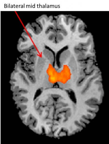
In the sleep study, the participants lowered the alpha frequency and raised the theta frequency with the help of an Aiji neurofeedback protocol in order to reach a state of deep relaxation (pre-sleep) while being scanned by the imaging device (Fmri - Functional MRI). In this study it was found that the middle part of the thalamus showed a decrease in activity both at the beginning of the transition and later in the transition to sleep. This finding supports the results of previous studies that attribute to the middle part of the thalamus a central role in regulating levels of awareness of external stimuli. That is, the thalamus functions as a gate and a navigator between the incoming sensory stimuli and the transition to the various brain systems that handle them, as suggested by Nicholas Schiff in an article from 2013 (is it really important to mention this?). Other brain systems that have been found and whose activity has increased are the systems that are related to the physiological physical changes. Studies of the process of anesthesia and the transition between wakefulness and sleep describe a variety of physiological changes such as decreased breathing rate, lower muscle tension, slow eye movements and visual and auditory hallucinations.
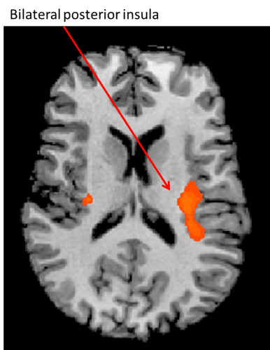
For example, in the study, the ventral tegmental area, which is a nucleus in the midbrain area, as well as the superior nucleus, were found, both of which are involved in eye movement and eye-head coordination. The frontal cerebellum also showed increased activity and findings from previous studies show that this region is associated with the control of autonomic responses related to breathing as well as the control of body stability. In the second part of the transition to sleep, activity increased in other areas such as the posterior insula and the medial posterior cingulate cortex. Together they form part of the posterior salience network, a network mentioned in William Shearer's (2012) article (is it important to mention this?) as being related to bodily awareness that regulates physiological responses and internal balance (homeostasis).
That is, in the process of transitioning to sleep, while brain areas that are attentive to signals coming from the outside show a decrease in activity, (activity increases in the areas that are attentive to signals coming from the inside.
The interesting finding from the study is that the two groups of subjects (those who managed to reach a state of deep relaxation and those who did not), showed similar activity in areas of the brain related to the control of physiological indicators (for example, lowering the breathing rate or relaxing the accompanying muscles during sleep), but only the group that managed to disconnect from the environment and transmit awareness from the outside Inside is the group that continued the process of transitioning to sleep.

One response
You mentioned that the 2 groups of subjects (the one that reached relaxation and the one that didn't) showed similar activity in areas of the brain related to the control of physiological indicators. That is, no objective difference was found between the 2 groups?
I read a study that used TMS in which increased brain flexibility in the cerebral cortex was discovered, among people with poor quality sleep: sleeplessness.org.il/insomnia/brain-differences-associated-insomnia