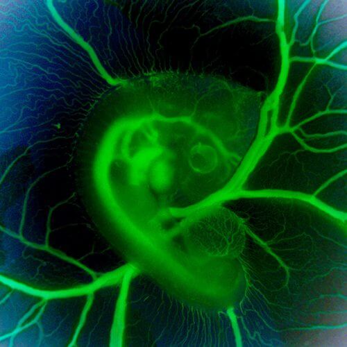
Videos documenting chicken embryos reveal the existence of special cells that play a crucial role in the development of the heart and blood vessels

The cardiovascular system is one of the first organ systems to form in the body. The reason for this is because the developing fetus needs blood vessels to transport the oxygen and nutrients necessary for its growth. In fact, the fact that this process begins so early in embryonic development contributes to the fact that scientists are still faced with many unanswered questions regarding the origin of the primitive heart and blood vessels. One of the main questions is: how and where are the progenitor cells, those first cells destined to be part of the cardiovascular system, formed?
Dr. Liad Zamir, former research student in the laboratory of Prof. Eldad Tzhor from the department of molecular cell biology at the Weizmann Institute, developed a method for imaging the earliest progenitor cells of the heart system - using videos that follow these cells in real time throughout the development of the embryo. Dr. Zamir studied the process in fertilized chicken eggs, where a complex network of blood vessels is formed inside the yolk sac for the purpose of feeding the embryo. The research findings were published recently In the online scientific journal eLife.
Collaborating with the laboratory of Prof. Richard Harvey, from the Victor Chang Research Institute and the University of New South Wales in Australia, Tzhor and Zamir focused on a gene called Nkx2-5. This gene encodes a transcription factor - a regulatory protein that controls the expression of entire sets of other genes; In this case, the genes involved in heart development. "When Nkx2-5 is silenced in the mouse embryo, the result is severe defects in the blood vessels and heart," says Tzhor. "But only recently has the gene also been linked to the development of blood vessels. The new study revealed that in addition to directing the development of the heart, Nkx2-5 plays a central role in the initial development of the early blood vessels, and in the formation of the first blood cells. This role does not depend on his roles in the heart."

While observing the early expression of Nkx2-5 in chicken embryos, and later in mice, the research team discovered the existence of progenitor cells called hemangioblasts. These cells are the source of the blood and of the vascular progenitor cells, the cells from which blood vessels will develop. These unique cells are created from the mesoderm, the middle layer of cells that appears in the developing embryo during the gastrulation stage. In the past, scientists have debated passionately about the very existence of hemangioblasts, and if they do exist, about their possible role.
In the videos, the researchers could see the hemangioblasts moving in the fetus and producing "blood islands". They were surprised to see that some of the hemangioblasts moved to the heart, where they formed colonies of blood stem cells. This finding helped to understand other studies, where it was found that the early heart has cells that help in the production of blood cells. The researchers identified such special Nkx2-5 cells in the heart and also in the aorta – one of the main blood vessels in the fetus – that it forms, where they appear to make new blood cells. Later during development, these specialized cells move to the liver, where they are the source of blood stem cell formation in the fetus.
To test the results obtained in chicken embryos, the researchers used genetic methods to mark cells in mouse embryos. They showed that a population of hemangioblasts expressing Nkx2-5 appears in the yolk sac in mice. This finding indicates that the origin of the cells that express Nkx2-5 and form the blood system has been preserved throughout evolution.
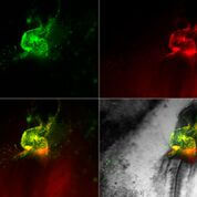
The development of the heart is in rapid motion. The early cells (in green and red) migrate towards the midline in the chicken embryo - where they join to form the embryonic heart
"20 years after the discovery of one of the main genes in the development of the heart, we were able to tell a new story about its activity," says Tzhor. "We showed that it acts for a short time at a very early stage in the development of blood vessels and the progenitor cells that produce blood - before its main activity in the heart cells. We presented solid evidence for the existence of these early cells, and for their contribution to the development of the heart and blood vessels."
Revealing the ancient origins of at least some of the cells that form the blood stem cells in the fetus, may help in the study of diseases that affect the cardiovascular system.
