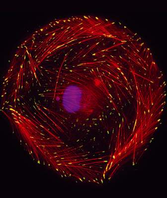
Adhesion of cells to their growth medium is not a simple and passive "adhesion", but rather a complicated process. It is done through "targeted adhesion areas", which connect the outer surface of the cell and the connective tissues

Adhesion of cells to their growth medium is not a simple and passive "adhesion", but rather a complicated process. It is done using "targeted adhesion areas", which connect the outer surface of the cell with the connective tissues. These adhesion areas consist of hundreds of different proteins, which connect the inner skeleton of the cell, indirectly, to the outer surface, and they play a central role in fixing the cell to its place in the tissue. Recently, it became clear that the contact areas also play a central role in sensing the properties of the cell's environment, and in transferring the information gathered inside, into the cell. A new study by scientists at the Weizmann Institute of Science reveals that the sensing processes controlled by cell adhesion are carefully controlled processes, so that even cells that migrate throughout the body usually "check the ground" before setting out on a journey, in order to prepare themselves for movement.
Prof. Benny Giger and Prof. Alexander Bershadsky, from the Department of Molecular Biology of the Cell, investigate the ways in which cells sense the physical properties of their environment, and react to them. Blood pressure, muscle tension, and stiffness and tension in tissues - all these are forces that act on cells, and influence their behavior and fate. In addition to the external forces, the cells themselves can also exert contraction forces through cellular fibers that belong to the "cytoskeleton": when a cell touches a surface, it loosely binds to it, while contracting, to check the surface while pulling. If the test shows that the surface contains the appropriate components for the cell, and has the appropriate mechanical properties (for example, softness or stiffness), the cell adheres to them, and in the process increases its adhesion sites, with the aim of creating focused adhesion areas, a process that causes it to spread over Surface.
To try to understand the complex interrelationships between the infected cell and the surface involved in the cell's sensing mechanism, Prof. Giger and Prof. Bershadsky, together with their research colleagues Dr. Masha Perger-Khotorsky and Dr. Alexandra Lichtenstein, designed and established an experimental system in which cells are positioned on polymeric surfaces that differ in their hardness, but are similar in their chemical properties.
A microscopic examination indicated great differences in the behavior of the cells on the two types of surfaces: the cells that were located on the softer surfaces spread out evenly in all directions, creating circular surfaces - similar to an "egg of an eye", while on the hard surfaces the cells took on elongated shapes. The elongated shape gives cells polarity - "head" and "tail", and is what allows cells to move and migrate - an essential feature for embryonic development, wound healing, and tissue growth and repair. A closer look also revealed significant differences in the structure of the targeted contact sites: on the soft surface, the round cells created many small adhesion points, which were uniformly distributed along the entire length of the cell's edge. In contrast, on the hard surfaces the cells created large focused contact areas, which tended to develop mainly in the future head and tail areas, even before the elongation process began. In other words, the choice of whether the cell will be elongated and ready for travel, or a round and stationary form, depends directly on the hardness of the surface on which the cell is located; The targeted contact areas act, among other things, as stiffness sensors.
To understand the cellular infection control process at the molecular level, the researchers systematically blocked about 80 different genes, which code for proteins that participate in signal transmission processes in the cell, using gene silencing technology. Then check how each of these cells reacts to the surface. At this stage, the scientists collaborated with Prof. Zvi Kam from the department of molecular biology of the cell at the institute, who assisted with automatic microscopy, automatic analysis of visual information, and the quantification of the data, and with Dr. Ramashwami Krishnan from Harvard University, who developed a method for measuring the forces created in cells, when they pull on a surface.
The analysis of the findings indicated that, in some cases, the silencing of the genes caused the cells to lose their ability to polarize, or to adjust the size of the targeted adhesions they create to the hardness of the surface. In other cells, gene silencing caused changes in the cell's grip on the surface, or the cell's ability to sense and respond to mechanical forces. The conclusion that emerged from the study - published in the journal Nature Cell Biology - was that cell polarization is an extremely complex process, driven by mechanical forces and mediated through targeted cell contacts. The control of the process occurs in several stages, which affect the production of forces in the cell, and also manage the response to the various forces. "The amount of control factors involved in this process surprised us," says Prof. Bershadsky. Prof. Giger concludes: "We discovered a strong connection between the development of cellular contacts with their 'living environment' and the production of force by the cells, their polarization and migration, and we identified some of the main control factors in this process."
These findings may be useful in many areas of biology and biomedical research. So, for example, the scientists believe that their insights are relevant for the cells that line the blood vessels. These cells are exposed to eddy currents and pressure changes - which may lead to the formation of arterial deposits or the abnormal development of peripheral blood vessels. A better understanding of the process may help identify possible therapeutic targets, which can be attacked in the future with drugs.
