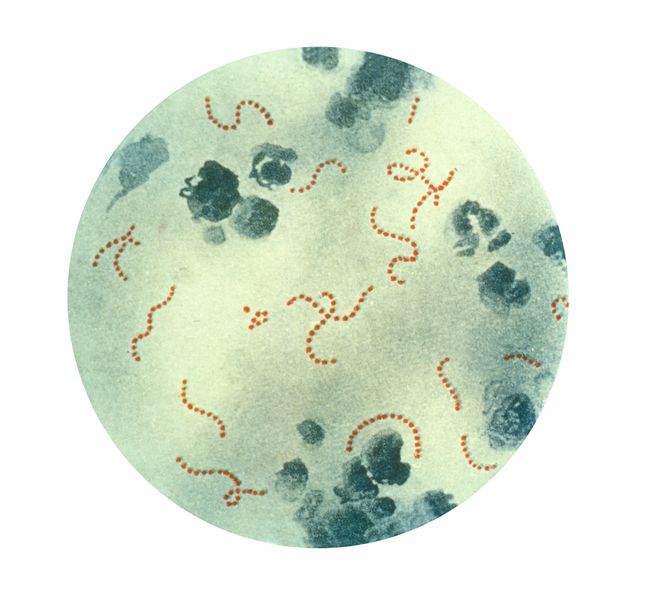
The bacterial cell wall, which is a target for possible antibiotics, such as penicillin, actually consists of a thin monolayer of sugar chains (carbohydrates) bound together by proteins, thus wrapping the bacteria like a belt around the waist

The bacterial cell wall, which is a target for possible antibiotics, such as penicillin, actually consists of a thin monolayer of sugar chains (carbohydrates) bound together by proteins, thus wrapping the bacteria like a belt around the waist.
This is according to a study conducted by a group of scientists from the California Institute of Technology (California Institute of Technology also known as Caltech). The first glimpse ever into the three-dimensional structure of the cell wall was made possible with the help of state-of-the-art microscopic methods that provided scientists with the ability to visualize these biological structures at nanometer levels. "This is both a technological and a biological advance," says scientist Grant Jensen, a professor of biology at the institute and the study's principal investigator.
"Bacterial cells rely on a cage-like network that surrounds them and maintains their integrity," explains the researcher. "Without the existence of this molecular sac - the bacterium would not survive; Most likely he would have disintegrated."
This sac, known as a sacculus, consists of peptidoglycan - a network of polymers consisting of long chains of polysaccharides linked together by short peptides. Peptidoglycan is the main structural component of the cell wall of bacteria, and it is what gives it its shape and strength.
Many antibiotic drugs, such as penicillin, interfere with the construction of peptidoglycan, and especially the action of transpeptidase, the enzyme that builds it, thus damaging the bacterial cell wall. Mutations that cause changes in the transpeptidase, and consequently interfere with the interactions between it and the antibiotic drug, are a significant factor in the development of antibiotic resistance.
Since the dawn of time, researchers have been interested in understanding the detailed and precise structure of this vesicle. In particular, the researcher and his colleagues wondered whether the sugar chains - linked by the short peptides - really wrap the cell like a belt or if they protrude outside the cell envelope like grass above the ground. The answer to this controversy eluded the scientists, because the attempt to predict such tiny biological details was beyond their technological reach. So far.
"Six years ago, a grant from the Moore Foundation allowed us to purchase what can be called the best electronic cryogenic microscope in the world," says the researcher. "This equipment allowed us to obtain a different type of photographs of tiny biological details that did not exist before. These photographs are XNUMXD images at molecular resolution - you can literally start visualizing individual biological compounds. With it we could observe this network of sugar chains. It was just amazing.”
By combining the microscopic photograph with a purification method consisting of removing the vesicle envelopes and flattening them in a thin layer of water, researcher Lu Gan, the first author of the article, was able to visualize the detailed three-dimensional structure of the peptidoglycan network and conduct a sort of simulated spatial "tour" of the bacterial vesicle.
"What we saw were long tiny tubes that wrapped around the bladder like a person's ribs or a belt around the waist," says the lead researcher. "We also saw that the vesicle consists of a thin monolayer."
"This is a clear answer to this ancient question," adds the researcher. "Today we know the exact details of the most basic compound that affects the spatial structure. Today we know what the correct answer is in the face of a wide spectrum of other answers, some of which are completely wrong."
Understanding the exact composition of the cell wall is extremely important, says the researcher, because for a very long time scientists have been in the dark about the most basic physical and mechanical aspects of bacterial life, including why they are built in the way they are. "It is difficult to properly understand how a building is put together unless you are able to see the building blocks themselves," he explains. "Now that we are able to see the building blocks - the most basic details in the structure of the vesicle - we are closer to understanding how the bacteria are able to control their growth and how drugs that prevent this can work."

4 תגובות
Hello. I saw your article on E_COLI. I am looking for cheap solutions to convert cellulose to monosaccharides.
A good article written in simple and understandable language. Interesting and disturbing.
the link is not working.
Too bad there is no picture. (The link to the original news doesn't work either)