Weizmann Institute scientists present a new method for imaging a single electron
For several decades now, MRI scans have been an essential means of identifying a multitude of diseases, and as a result, saving countless lives. Simply put, this technology makes it possible to produce a coherent image of the inside of the body, by measuring the density of water molecules. The image is obtained with a resolution of 1 cubic millimeter - an excellent degree of separation if our aim is to check if there is, for example, a tear in the meniscus in the knee. But what about the structure of a single molecule? The size of such a molecule is around 5 cubic nanometers - that is, it is about 10 trillion times smaller than the highest resolution that the most modern MRI scanner is capable of reaching, so - on the face of it - this is an impossible task. But for Dr Amit Finkler From the Department of Chemical and Biological Physics at the Weizmann Institute of Science - this is the mission. In the article thatRecently published, Dr. Finkler, research student Dan Yudilevich and their partners from the University of Stuttgart, Germany - Dr. Rainer Stöhr and Dr. Andrey Denisenko - succeeded in completing another step in this direction, when they demonstrated a new method for imaging single electrons. Although the method is in the initial development stage, it is possible that in the future it will be applied for imaging at the molecular level - which may revolutionize the development of new drugs and the characterization of quantum materials.

MRI was and still is a breakthrough method, but in some cases it has drawbacks. For example, in order for it to work properly, a relatively large sample is needed - at least a few hundred billion water molecules - and as a result the displayed output is a kind of average of smaller components - which the machine is unable to recognize. In most types of diagnoses - the average is actually the desired and optimal result. But in other cases - for example, in drug development - work is required on as small a sheet as possible, which will provide accurate results. Beyond that, when averaging so many different components - some of the information may fall through the cracks, thus even preventing the discovery of important processes that happen on an extremely small scale. For this purpose, the scientists propose molecular close-up photography.
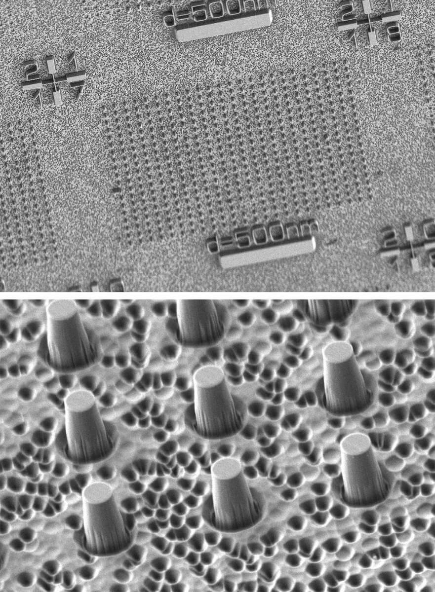
The new method can locate the exact position of a single electron by applying a magnetic field and rotating it around a synthetic diamond, which is used as a quantum sensor
What would a more accurate imaging device look like, capable of working on an extremely small scale? Dr. Finkler, Yudilevich and their partners developed a method that can locate the exact position of a single electron by applying a magnetic field and rotating the field around a synthetic diamond, which is used as a quantum sensor. In fact, it is not the diamond itself acting as a sensor, but a tiny defect in it - the size of a single atom - called a "nitrogen-vacancy center". Due to the atomic size of the sensor, it is specially adapted to distinguish the changes that occur near it, and due to its quantum nature, it can distinguish between a single electron and a cluster of electrons. Due to these properties, it is particularly suitable for measuring the position in space of a single electron with an extraordinary level of precision, which provides us with a glimpse of the imaging technologies of the future.
"It will be possible to use the new method we developed," says Dr. Finkler, "to provide doctors and scientists with an additional and complementary point of view - to the existing imaging methods." The development of such a tool may allow scientists to closely study the structure of important molecules, thus paving the way for new discoveries. The scientists envision a future where we can use the new method to image a wide variety of molecules, which will hopefully reveal more layers of the holy molecular trinity: structure, function and interactions.
Multidisciplinary
Dr. Amit Finkler first came to the Weizmann Institute of Science as a bachelor's graduate. He completed his second and third degrees at the Institute in the laboratory of Prof. Eli Zeldov, in the Faculty of Physics, before going to the University of Stuttgart - where he did his post-doctoral research. Five years ago he returned to Rehovot and established his own laboratory - in the Faculty of Chemistry.
"I'm a physicist," says Dr. Finkler, "but working in the Faculty of Chemistry creates unique opportunities for the research we carry out in my laboratory. We have physicists, but there are also chemists and engineers. It's an interesting combination, and the possibility to combine different research methods and disciplines is necessary for the type of science I'm interested in. I like to adjust lenses, I enjoy cryogenic burns, and now I spray engineered molecules on our diamonds."
More of the topic in Hayadan:
