Hebrew University researchers used for the first time a method that has existed for many years to reveal the connectivity patterns in the brain at the cellular level, and determined that the application of the method has great potential in understanding pathological conditions in the brain. The research is published in Science

The human brain is buzzing with activity - the 86 billion nerve cells (neurons) that make it up are constantly sending electrical signals to each other. These nerve signals are sent along a system of fibers known as the "white matter", and are the basis of all brain functions. Mapping the fibers that connect the different brain regions is an ongoing challenge in neuroscience. The currently existing methods for mapping the white matter are limited to research in animals, or require advanced equipment for data collection and processing.
Recently, a new approach to brain nerve fiber mapping was developed by Prof. Aviv Metzer and Dr. Roi Shor from the Edmond and Lily Safra Neuroscience Center (ELSC) at the Hebrew University of Jerusalem, which was published today (Thursday) in the international journal Science. The team of researchers used a common technique that has been around for almost 140 years, but has never been used to study connectivity in the brain. It is based on brain cell staining known as Nissl staining, which was developed in the eighties of the nineteenth century by the German researcher Franz Nissl. This method, in which the cells are colored in an oval color, has been used for over a century in the study of neurons in the brain, and its use has revolutionized the way we understand the structure of the cerebral cortex (the outer gray layer of the brain). however, The Hebrew University team is the first to use Nissl staining to reveal the fiber pathways in the white matter of the brain.
The white matter consists mainly of fibers called axons (nerve fiber) and cells called glial cells (non-neuronal support cells for neurons, which provide protection to the brain nerves and hold the nerve cells in place). Until recent years, glial cells did not receive much attention in research, because their role in the brain was thought to be marginal. In fact, the name "Galiya" is derived from ancient Greek, and means "glue".
Several years ago, Dr. Shor joined Prof. Metzer's laboratory as a doctoral student, and decided to look for images of brain tissue stained with the Nissel method. "It just intrigued me," recalls Dr. Shor, "the textbooks are full of illustrations, but I wanted to understand how the white matter fibers of the brain look like in reality." To his surprise, he noticed that the glial cells formed a pattern of short rows. Furthermore, it appears that the glial rows were arranged along the local nerve fibers. "We conducted a literature review to see if anyone else had noticed this," Dr. Shore shared, "And we came across an article from 1992 that showed that the glial cells were indeed found to be organized in short rows along the nerve fibers. This article has not received the attention it deserves.'
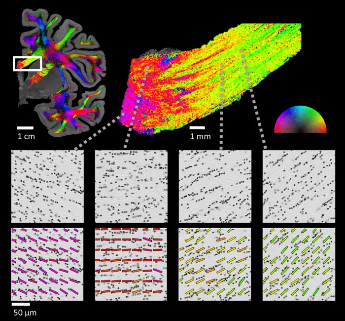
NISSL imaging. Courtesy of the Hebrew University
Shore took time off to engage in other research, but recently returned to his work on these cells. In the current work, the researchers realized that by means of simple computational tools from the field of image processing, they would be able to take advantage of the spatial arrangement of the glial cells and deduce the direction of the fiber pathways in the white matter. "As soon as I saw the spectacular pictures of theNissl-ST I understood that we found a treasure, no less than that, that researchers have been looking for for years," shares Professor Metzer and adds: "We found a simple method to identify the structural description of the white matter at the cellular level."
In conclusion, according to the researchers, many brain disorders are linked to changes in neural connectivity, so the application of Nissl-ST has great potential for use in future studies of white matter in normal development, normal aging and pathological conditions that affect the white matter in the brain, such as in neurodegenerative diseases and schizophrenia.
