If it were possible, for example, to mark with colors the expression of genes in the cells of the body, vital biological processes that are now hidden from view would unfold before our eyes. The fluorescence revolution makes it possible to illuminate, in the full sense of the word, the activity occurring in the cells, but it does not allow observing the processes occurring in the depths of the body
Even die-hard fans of black-and-white films cannot deny that the introduction of shot color photography has given new life to the art of cinema. And as far as the documentation of what happens in our body is concerned, there is no substitute for color. If it were possible, for example, to mark with colors the expression of genes in the cells of the body, vital biological processes that are now hidden from view would unfold before our eyes. The fluorescence revolution makes it possible to illuminate, in the full sense of the word, the activity taking place in the cells, but it does not allow observing the processes taking place in the depths of the body, since the tissues in the way dim the glow of the fluorescent proteins. In a new study published today in the scientific journal Nature Biotechnology, The scientists of the Weizmann Institute of Science managed to penetrate through the tissues in a roundabout way: they "injected" new life into the grayish scans of magnetic resonance imaging (MRI) and followed with colors the expression of two different "reporter genes" (reporter genes) at the same time. "In the future, MRI scans may be able to replace invasive tests or removing samples from the body for examination under the microscope," outlines the head of the research team, Dr. Amnon Bar-Shir, the horizon opened thanks to the new discovery.
When the 2008 Nobel Prize in Chemistry was awarded for the development of fluorescent proteins, one of the winners, the late Roger Tessien, admitted that the method had limitations, and that in the future it might be possible to overcome them with the help of other technologies such as MRI. The vision outlined by Tessien is based on the fact that, unlike light waves, the radio waves on which the MRI method is based, are not blocked by body tissues, even when it comes to particularly thick tissues. However, the MRI scans we know today - in black and white - are mainly adapted to imaging structural elements, and the advanced ones, which allow color coding, were not adapted to mapping gene activity in cells. Even when the MRI was previously adapted to monitor gene expression, it was possible to monitor only one gene at any given time - because its activity was marked by a dark dot on a black-and-white background. Therefore, the ability to monitor several genes that are expressed at the same time - preferably with the help of different colors, such as fluorescent labeling - may represent a significant leap forward in the field of imaging.
Dr. Bar-Shir's group in the Department of Molecular Chemistry and Materials Science decided to pick up the gauntlet thrown by Tessien. The research team led by faculty scientist Dr. Hila Elosh-Arnon, in collaboration with Prof. Sheral Fleishman And Dr. Olga Khersonsky from the Department of Biomolecular Sciences, developed a two-step method: in the first step, the researchers created two groups of genetically modified cells, with each group designed to express one of two designated proteins. At the same time, they created two types of molecular detectors designed to move through the bloodstream and accumulate only in those cells that expressed the engineered proteins. The detectors were designed to operate at separate frequencies and cause different colors to appear in MRI scans.
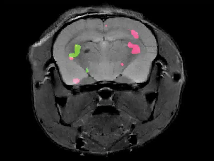
After the preparations were completed, the researchers applied the method to laboratory mice that were inserted into one of the most powerful MRI machines operating in the world today. The two glowing colors did not take long to appear: the resulting scans revealed precisely the location of the cells that express the two genetically engineered proteins in the mice's brains. In doing so, the researchers demonstrated the feasibility of monitoring the expression of various genes in the depths of the brain and body using MRI. Further studies will aim to develop the method to map a larger number of genes simultaneously.
This new imaging method paves the way for the use of MRI to monitor a wide variety of biological processes in the living body for research purposes - and in the future for medical purposes as well. For example, this method may allow researchers to track how one brain area affects another. If and when the method is adapted for use in humans, it may even allow researchers and doctors to replace invasive tests and procedures. For example, it will be possible to monitor innovative cancer treatments that include the injection of cells and simultaneously monitor the injected cells and the tumor cells.
Nishant Tirokoti, a research student in Dr. Bar-Shir's laboratory, also participated in the study; Dr. Yoav Peleg, Dr. Orli Dim, Dr. Shira Albek, Dr. Alexander Brandis and Tebi Melman from the Department of Life Science Research Infrastructures; Dr. Liat Avram and Dr. Talia Harris from the Department of Chemical Research Infrastructures; and Dr. Nirbhai Yadav from Johns Hopkins University.
More of the topic in Hayadan:
