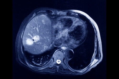
The method developed at Stanford could reduce patients' risk of developing secondary cancers later in life. The new method is a different version of the imaging method based on magnetic resonance (MRI) and it uses new contrast materials to locate the tumors.

A method has been developed for scanning the bodies of young cancer patients to detect tumors without exposing them to radiation. The method could reduce patients' risk of developing secondary cancers later in life. The new method is a different version of the imaging method based on magnetic resonance (MRI) and it uses new contrast materials to locate the tumors. The study found that the new method is just as effective as other scanning methods based on ionizing radiation designed to locate tumors, especially positron emission tomography (PET) and computed tomography (CT).
Researchers from the Stanford University School of Medicine and the University Children's Hospital have developed a method for scanning the bodies of young cancer patients to locate tumors without exposing them to radiation. The new method could reduce patients' risk of developing secondary cancers later in life. The new method, described in the scientific journal The Lancet Oncology, is a slightly different version of the MRI method and uses innovative contrast agents to locate tumors.
Despite the fact that the technology of a full body scan using PET-CT provides essential information for the detection of cancer, it has one major drawback: a single scan exposes the patient to radiation equivalent to 700 x-rays. Such a high level of exposure is especially dangerous for children and young boys who are more vulnerable to radiation than adults due to the fact that they are still growing. In addition, it is likely that the children will live long enough that they may develop a secondary cancer. "I'm pretty excited about an imaging test for cancer patients that requires zero radiation exposure," said one of the researchers. "This is an important and welcome matter."
The research team compared the improved MRI method with the standard PET-CT method among 22 patients aged 33-8 who were patients with lymphoma or sarcoma. These types of cancer originate in the immune system and bone, respectively. Both of these types of cancer can spread through the bone marrow, lymph nodes, liver and spleen.
In the past, several hurdles prevented doctors from using whole-body MRI to detect abnormalities. The duration of the scan itself lasts two whole hours. In contrast, a full body scan with PET-CT takes only a few minutes. And more importantly, with the MRI method it is not possible to differentiate between cancerous and healthy tissues in many organs. In addition, existing contrast agents - the same chemicals injected into the body to "paint" the tumors - are released from the tissues too quickly to allow a scan that lasts two hours.
In order to locate tumors with the help of the MRI device, the research team used a new contrast agent containing iron nanoparticles. The injection of these iron nanoparticles is approved by the US Food and Drug Administration for the treatment of anemia, and the researchers obtained the administrator's approval for the experiment in question. These nanoparticles remain in the body for many days. When used in the MRI machine, these particles make the blood vessels appear clearer, allowing accurate reception of the anatomical contours of the body. The nanoparticles also cause the bone marrow, lymph nodes, liver and spleen to appear black, making the tumors stand out even more.
The images obtained from the MRI device experiments provided information comparable in quality to the information obtained from the PET-CT device scans that the test patients had as part of their treatment. With the PET-CT method, 163 of the 174 tumors were located in 22 patients; Using the MRI method, 158 of the 174 tumors were located. Both methods have similar levels of sensitivity, selectivity and diagnostic accuracy. "We were able to find a new way to combine anatomical and physiological information obtained with MRI and make it more efficient," said the paper's lead author.
None of the patients experienced side effects in response to the iron nanoparticles, although the US Food and Drug Administration previously stated that there is a low risk of an allergic reaction to the coating of the nanoparticles. Radiology specialists at several academic hospitals are looking for ways to reduce the amount of radiation to which children are exposed, explains one of the researchers, who also states that she shares the details and results of the new method with colleagues from around the country.
"A certain type of whole-body MRI imaging test is already available in many of the major children's hospitals," says the researcher, noting that in order for such a method to become widespread, doctors want to be convinced that it really works. "The method is slowly entering clinical use, but the doctors are cautious and interested in proof first," she notes.
Another study is planned for the validation of the new method among wider and more diverse groups of cancer patients, and for examining the possibility of using the method to monitor tumors during cancer treatment. In addition, the method will also be useful and important for examining the patients at the end of their treatment when the ability to monitor their condition without a scan is a valuable ability.
The news about the study

3 תגובות
Important information (by the way, the link to the study below leads to the photo site and not to the study).
The MRI actually started with a patent that Damdian issued in 1971 for an idea for a scanner that would be used for whole-body scanning (the machine was then called "made without fear"). It is important to note that an MRI scan for two hours is not less risky and it also damages the DNA in its own way (although the effect of the damage is not yet clear, certainly not as clear when it comes to ionizing radiation).
Let them find a cure for this damn disease (on any issue).