Engineers have succeeded in developing an atomic force microscope that scans images at a rate 2000 times faster than commercial models sold on the market. Using this innovative high-speed tool, the researchers were able to produce images of chemical processes occurring on the nanometer scale at a speed approaching the level of real-time video.
[Translation by Dr. Nachmani Moshe]
Engineers have succeeded in developing an atomic force microscope that scans images at a rate 2000 times faster than commercial models sold on the market. Using this innovative high-speed tool, the researchers were able to produce images of chemical processes occurring on the nanometer scale at a speed approaching the level of real-time video.
Advanced atomic force microscopes (AFMs) are designed to produce images of tiny structures with the size of fractions of a nanometer - a million times smaller than the thickness of a human hair. In recent years, microscopes of this type have been used to photograph single-atom structures, such as single strands of DNA and even separate hydrogen bonds between molecules. At the same time, scanning such images is a meticulous operation that requires a lot of time. Therefore, microscopes of this type are mainly used for examining stationary samples since their activity rate is too small to capture active and changing environments.
Now, engineers from the Massachusetts Institute of Technology (MIT) have succeeded in developing an atomic force microscope that scans images at a rate 2000 times faster than commercial models sold on the market. Using this innovative high-speed tool, the researchers were able to produce images of chemical processes occurring on the nanometer scale at a speed approaching the level of real-time video. In one demonstration of this device's capabilities, the researchers scanned a 70 x 70 micron sample of calcium carbonate while immersed in deionized water and then exposed to sulfuric acid. The researchers were able to see the reaction during which the acid corrodes the calcium carbonate while enlarging the nanometer holes inside it in the process of "peeling" the crystalline pattern of the material layer by layer, in just a few seconds. The lead researcher, Professor Kamal Youcef-Toumi, says that the sensitivity and speed of the device will allow scientists to watch atomic processes like in high-resolution "videos". "The researchers will be able to see, for example, reactions such as condensation, deficit, dissolution or sedimentation of various substances, and the way in which these reactions occur in real time - processes that have never been seen at such a resolution." The research findings have long been published in the scientific journal Ultramicroscopy.
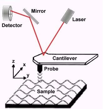
An atomic force microscope scans samples with an extremely tiny detector, or needle-like tip, that moves slightly above the surface of the sample, tracking its topology, similar to how a blind person reads Braille. The sample is placed on a moving platform, or scanner, which moves the sample along and under the detector. In light of the fact that a microscope of this type includes extremely tiny structures, the device must operate slowly, layer by layer, in order to avoid any sudden movement that could change the composition of the sample or blur the resulting image. "If the sample is stationary, then the image can be obtained within 10 minutes of slow scanning," explains the lead researcher. However, if the sample is moving, then this mechanism is not suitable. In order to speed up the scanning process, the scientists are trying to build tiny, sharper substrates that are able to scan the samples more quickly, even though the scanned area is smaller. The researcher explains that although scanners of this type work quickly, they do not allow researchers to quickly switch to remote photography in order to see a wider picture or examine a larger number of details in the image.
The researcher came up with a design that would allow simultaneous high-speed scanning of both a tiny area and a large area. The innovation of the device lies in a multi-functional scanner and its control: the substrate on which the sample is placed includes both a tiny and fast scanner and a larger and slower scanner, which work together as one system to obtain a high-speed XNUMXD scan. Previous attempts to get a multi-operational scanner were doomed to failure due to the interplay between the two scanners: the movement of one scanner could impair the accuracy and smoothness of the second scanner. Also, the researchers discovered that the ability to control each of the scanners separately is problematic. The lead researcher explains that until today the scientists were required to make many adjustments and adjustments to the device's components in order to perform a double scan. In order to simplify the control of the dual scanner, the researcher developed algorithms that take into account the effects that each of the scanners has on the other. "Our controller is able to move the tiny scanner in a way that does not vibrate the larger scanner, this is because we know in advance what types of movements will cause this, and vice versa - that is, which movements in the large scanner will cause an effect on the tiny scanner." "In the end, the two scanners work in sync and for the researcher it is a single high-speed scanner that does not add any complexity to the operation of the device."

One response
Innovative until it reaches the computer.
WIN XP SHULTATTAT.