Inspired by the "Body Worlds" exhibition: a method that promises to speed up biomedical research
A few years ago, Viviana Gradinaro was cutting thin slices of mouse brain in a neurobiology laboratory. From the XNUMXD results of this layout she slowly assembled a XNUMXD computer model. In her free time she used to visit the "Body Worlds" exhibition. The thing that fascinated her the most was the display of the human circulatory system, which was produced from blood vessels that had undergone a process that made them look like plastic. Then the idea occurred to her that a similar process would make it possible to do most of the things she does in the lab more efficiently.
Methods for "tissue shading" have been accepted for more than a century, but they involve soaking the tissues in solvents. This is a slow process that usually destroys the fluorescent proteins necessary to mark cells of interest. Gradinaru, then a doctoral student, and her colleagues in the laboratory of the late neuroimmunologist Paul Patterson set out to create a better method. To this end, they focused on replacing the fat molecules that make the tissue opaque to light. But to prevent the tissue from collapsing, they had to find a substitute that would keep the shape given to it by the fats.
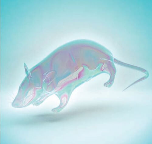
In the first step, they injected formalin into the body of a mouse that had just been killed, through its heart. They then stripped his skin and filled his blood vessels with acrylamide monomers, white, odorless crystals. The monomers connected to each other and formed a hydrogel network that replaced the lipids and made the tissue clear. Within two weeks the whole body of the mouse became transparent.
Before long they were using transparent mice to map their entire nervous system. The transparency allowed them to identify nodes in the peripheral nervous system: tiny associations of nerve cells about which knowledge is limited. They mapped the spread of a virus that crossed the blood-brain barrier. They did this by marking the viruses with a fluorescent substance, injecting them into the rodent's tail and watching them reach the brain. "It's like looking at the whole world instead of its individual pieces," says Gradinaro. The process reduces the chance of human error, speeds up laboratory work, yields richer data and requires fewer experimental animals. Gradinaru sends the prescription for its hydrogel solution to any laboratory that requests it. Her next task is to use the method to find, map and study cancer tumors and stem cells.
The article was published with the permission of Scientific American Israel
