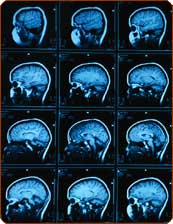
Innovations in the field of imaging allow scientists to verify what is happening in the brain in a series of thought processes and more. The method will allow neuroscientists to create architectural maps of brain functions, such as vision, movement and learning at a single-cell level of precision
Michael Speer, Scientific American
Neuroscientists explore areas that have not yet been mapped in the microscopic world of nerve cells (neurons). They observe the brain cells in action, discover microscopic evidence of Alzheimer's in the living brain and even do a bit of mind reading. The 90s were dubbed "the decade of the brain", but scientists in the XNUMXs are examining the living brain with much more wondrous precision.
To study the function of neurons on a microscopic scale, researchers usually use thin glass electrodes, but this method cannot provide the exact location of the cells in a living animal. The researcher can place the cell accurately only by injecting a chemical marker that can be seen under a microscope, and this only after the animal is killed.
A new technique - called functional imaging of a single neuron - now allows scientists to observe brain cells in action while they are still inside the brain. Already a year ago, R. Clay Reed and his colleagues from Harvard Medical School used a laser and a microscope to produce time-lapse images that recorded the activity of hundreds of neurons simultaneously in the visual cortex of laboratory cats and laboratory rats. The method, described in Nature in February 2005, will allow neuroscientists to create architectural maps of brain functions, such as vision, movement and learning at a single-cell level of precision.
Neuroscientists are not only building better maps of the brain, but they are also moving towards reading minds. Yukliaso Kamitani of ATR Computational Neuroscience Laboratories in Japan and Frank Tong of Vanderbilt University have shown that there is a close coupling between brain states, as measured by functional magnetic resonance imaging (fMRI), and subjective mental states.
In an article published in the journal Nature Neuroscience, the scientists described how they were able to predict which of eight graphic patterns the subject was looking at. They did this by deciphering the activity of a small group of neurons in the cerebral cortex responsible for vision. The scientists believe that it is possible to expand this method of mind reading and use it to study the neural basis of cognition, memory and other types of "thought content". It's all well and good to observe the brain doing its job when everything is fine, but when a disease breaks out, scientists want to look inside to find what the possible cause of the problem is. Neuroscientist Bradley Hyman from Massachusetts General Hospital and his colleagues have developed tools that can track microscopic neuronal changes in a live mouse used as a model for Alzheimer's disease. Using a multi-photon microscope and a fluorescent tracer, the scientists were able to distinguish with microscopic precision the plaques of amyloid - the hallmark of the disease. The scientists are now testing a similar method that uses positron emission tomography (PET) to diagnose and study the progression of the disease in humans.
These new techniques allow scientists to glimpse what's going on in the brain, but a more recent development will help them understand what they're seeing. In the April 22 issue of the journal Physical Review Letters, Nathan N. Urban of Carnegie Mellon University and his colleagues described a method that allows them to predict how groups of neurons synchronize their activity. Synchronized activity is the basis for encoding information and storing it in the brain, so their research has broad implications for clarifying the way the brain does the amazing thing called thinking.
