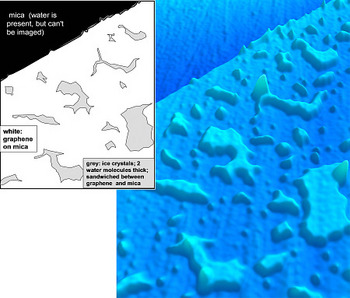
Researchers from the California Institute of Technology (Caltech) have developed an innovative method - using a one-atom-thick sheet of carbon atoms - to observe the atomic structure of particles. The method, which was used to produce the first ever direct image of how water coats surfaces at room temperature, can also be used to obtain the images of other particles, including antibodies and various bio-particles.

An article, describing the method and the experiments in the water layers, appears in the prestigious scientific journal Science.
"Almost all surfaces contain a coating of water on them," says researcher James Heath, professor of chemistry at Caltech, "and these waters are responsible for the properties of the interactions with the environment close to them." Although surfaces covered with water are common, their study is extremely problematic since the water droplets are "in constant flow, and do not settle at a certain point long enough to measure them," explains the researcher.
Quite by chance, the team of researchers developed a method to immobilize the moving mules, under room temperature conditions. "It was a happy randomness - one that we were wise enough to notice its importance," notes the researcher. "We studied graphene that was placed on a flat surface of mica (mica, a transparent mineral) and discovered a number of island-like nanostructures trapped between the graphene layer and the mica layer that we did not expect to get."
Graphene, which consists of a monoatomic layer of carbon atoms in a honeycomb lattice structure, should be completely flat when placed on a smooth surface. The researchers hypothesized that the unusual structures might be water, trapped under the graphene; Water frogs, after all, are everywhere.
In order to test their hypothesis, the researchers performed additional experiments in which they injected varying amounts of moisture into the graphene sheets. The strange structures became more common in high humidity, and disappeared completely in dry conditions, leading the researchers to conclude that they were indeed water droplets hiding under the graphene sheets.
At this stage, the researchers realized that the graphene sheet is a kind of "pressure wrap" that tightly binds the water particles, until their individual atomic structure could be discerned using an Atomic Force Microscope (AFM).
"The method is completely simple - it's really a miracle that it works," notes the researcher. The method, he explains, "is similar to the method in which carbon or gold atoms are injected into the interior of biological cells in order to simulate them - the carbon or gold atoms fix the cells. Here, the graphene completely covers the water particles weakly adsorbed to the surface and fixes them in place, for a period of at least several months."
Using the method, the researchers discovered new details about how water coats surfaces. They found that the first layer of water due to the mica actually consists of a layer the thickness of two parts of water, and has the structure of ice. Once the layer is fully formed, a second layer of double-thick ice is formed above it. Above this layer, "drops are obtained," explains the researcher. "It is absolutely amazing that the first two adsorbed layers of water form microscopic ice-like islands at room temperature," explains the lead researcher. "These fascinating structures are probably important in determining the surface properties of solids, in general, for example - their levels of lubrication, adhesion and digestibility."
Since then, the researchers have successfully tested other particles using flat atomic surfaces of various types - and this flatness is necessary so that the particles do not get stuck at the defect points on the surface, while losing the structure measured through the graphene layer. "We have not yet encountered a system in which this phenomenon does not occur," adds the researcher. The researchers are now working to improve the resolution of the method so that it can be used to image the atomic structures of bio-particles such as antibodies and other proteins. "In the past, we observed individual atoms of graphene using a Scanning Tunneling Microscope (STM), adds the researcher. "Separation capacity at a similar level should be achievable for graphene-coated foils as well."
"We can coat graphene on biological animals - including animals that are in a partially aquatic environment, since the presence of water does not interfere - and obtain their three-dimensional structure," the researcher notes. It may even be possible to determine the structure of complex systems, such as protein-protein conjugates, "which are extremely difficult to formulate," he says.
While the information obtained from one particle reveals its general structure, information from ten particles will reveal clearer characteristics - and a computerized collection of many identical particles can reveal every atomic hole and crack.

2 תגובות
sounds interesting. Is it possible to use the method to identify an exact structure of biological molecules similar to the ribosomes of Ada Yonat? Could this be an alternative or complementary method to the method she was working on? (I remember something related to crystals)
Wow! Buildings of complexes? Without breaking a headache about formation for crystallography? A real coup.