Scientists have been able to demonstrate how a method works to create XNUMXD images of biological structures under normal conditions while obtaining a resolution much higher than that of other existing methods. The method could help to understand how cells communicate with each other and provide important insights to engineers who develop artificial organs, such as skin and heart tissue
![Scientists from the University of Texas have succeeded in developing an innovative microscopic method for three-dimensional imaging of nanoscale biological structures, a method that is analogous to using a luminous rubber ball to obtain an image of a chair in a dark room. [Courtesy of Jenna Luecke]](https://www.hayadan.org.il/images/content3/2016/09/chair_illustration_final_830x4981-500x300.jpg)
[Translation by Dr. Nachmani Moshe]
Scientists from the University of Texas were able to demonstrate how a method works to create XNUMXD images of biological structures under normal conditions while obtaining a much higher resolution than other existing methods. The method could help to understand how cells communicate with each other and provide important insights to engineers developing artificial organs, such as skin and heart tissue. The research findings were published in the scientific journal Nature Communications.
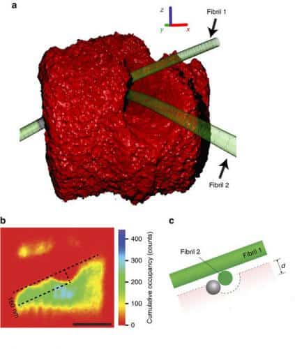
The scientists, led by the physicist Ernst-Ludwig Florin, used their innovative method, known as 'thermal noise imaging', in order to obtain images at the nanometer level of collagen fiber networks, which are part of the connective tissue found in the skin of . Examining the collagen fibers on such a scale allows scientists to measure, for the first time ever, main characteristics that affect the elasticity of the skin, findings that could lead to the improvement of the designs of artificial skin tissues. Obtaining clear XNUMXD images of nanoscale structures in biological samples is a difficult challenge, in part because of the tendency of the biological structures to be flexible and immersed in a liquid solution. This means that tiny changes in heat cause structures to move back and forth, a result known as 'Brownian motion'. In order to overcome the blur in the image that this result creates, other imaging methods are often used that aim to correct the resulting images by adding chemicals that lead to the hardening of various structures, however, in these cases, the materials lose their natural and original mechanical properties. Scientists can sometimes correct the blur if they focus on rigid structures associated with surfaces, such as glass, but this measurement limits the types of structures that can be studied.

The scientists took a different approach. In order to obtain an image, they added nanospheres - nanometer-sized beads that reflect laser radiation from their surface - to the biological samples under natural conditions, fired a laser beam at the sample while obtaining super-fast photographs of the nanospheres as they are seen through a normal light microscope. The scientists explain that their method, simulating thermal noise, works like this: imagine you need to take a XNUMXD image of a room in total darkness. If you throw a glowing rubber ball into a room and use a camera to collect a series of super-fast shots of the ball being thrown across the room, you will notice that it is unable to pass through solid objects such as tables and chairs. A combination of millions of such images collected in this way will make it possible to create an image of the objects in the dark room (the points in space where the bullet did not pass) and of the rest of the space where there are no objects (the points in space where the bullet did manage to pass). In a thermal noise imaging method, the alternative to the rubber ball is the nano-ball that moves through space in a natural Brownian motion. "These chaotic fluctuations are a serious nuisance in most microscopy methods since they cause the image to blur," says the lead researcher. "On the other hand, we used this chaos to our advantage. In addition, we do not need to develop a complex mechanism to move our detector. We sit comfortably and let nature do its thing."
The original idea for a thermal noise imaging method was published and patented as early as 2001, but technical challenges have prevented it, until today, from becoming a fully functional applied process. The new means allows researchers to measure, for the first time ever, the mechanical properties of collagen fibers in biological systems. Collagen is a biopolymer that creates scaffolds for cells in the skin and contributes to skin elasticity. Scientists are still not sure how the collagen network gives rise to the elasticity of the skin, an important question that requires an answer if we want to develop properly functioning artificial skin tissues. "If you want to create artificial skin, you must understand the interactions between the natural components," said the researcher. "In the next step, it will be possible to design a better collagen network that functions as a scaffold that encourages cells to grow properly."
