A team led by an Israeli researcher created a XNUMXD tissue from embryonic stem cells for the first time
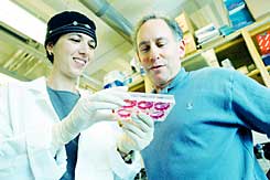
Dr. Shulamit Levenberg and Prof. Robert Langer, in whose laboratory the research was conducted. "The entire structure looks like a sponge in which the cells grow," says Levenberg. Photo: Donna Quinney, MIT
The severe shortage of transplantable organs has long spurred scientists to find alternatives to transplanting whole organs. The hope is that embryonic stem cells can do the job. Today, researchers are trying to grow embryonic stem cells in the laboratory, add various growth factors to them and chart their development into nerve cells, muscle cells, heart cells, insulin-secreting cells and more. However, in tissue, the cells are arranged in an organized structure and maintain mutual relations with each other. Therefore, if you want to create tissue from the embryonic cells, it is not enough to direct their differentiation, but you must ensure that they organize together in a three-dimensional array and function as a single unit.
In this context, the field of tissue engineering from stem cells has developed in recent years. The idea is to let the stem cells organize themselves on top of a biodegradable scaffold, made up of biological materials. The stem cells will gradually organize themselves on top of the scaffold and produce tissue, and at the same time the scaffold will gradually decay and the finished tissue will take its place. For this purpose, a specific cocktail of growth factors must be put together, which will lead to the differentiation of the embryonic stem cells into the desired tissue, a biological scaffold must be developed from materials whose rate of degradation matches the rate of differentiation and organization of the stem cells, and ultimately check if this prescription actually results in the creation of a functioning tissue. This is a complicated process, since the tissue consists not only of the cells responsible for its functions, but also of blood vessels that supply it with oxygen and nutrients. To date, researchers have not been able to create functional XNUMXD tissue from embryonic stem cells.
A first attempt in this direction has now been crowned with success. Dr. Shulamit Levenberg and a team of researchers from the Massachusetts Institute of Technology (MIT) succeeded in creating an organized XNUMXD tissue from embryonic stem cells grown on a biodegradable scaffold; The study was published at the end of October in the journal "National Academy of Sciences." Proceedings of the Levenberg, who will join the faculty of the Department of Biomedical Engineering at the Technion next year, conducted the research in the laboratory of Prof. Robert Langer. The team of researchers also included Prof. Yosef Itzkovits from the Faculty of Medicine at the Technion.
Levenberg initially focused on creating blood vessels outside the body. "The thought was that human embryonic stem cells could be a good source for this," she says. "A year ago we showed that it is possible to direct these stem cells to differentiate into endothelial cells, which line the inner wall of blood vessels; These cells will later produce tubes. When we let the human endothelial cells organize themselves on XNUMXD polymers and transplanted them into mice lacking an immune system (to prevent rejection), we saw that the resulting tubes, which were of human origin, integrated into the blood vessels of the mouse and that the blood cells of the mice wandered within them."
The next idea was to create a XNUMXD human tissue using the same method. "Complex tissue, for example heart tissue, includes muscle tissue, connective tissue, blood vessels and other factors that organize together to form the heart tissue as a whole," Levenberg says. "The hypothesis was that if we put the embryonic cells on a XNUMXD structure and give them signals that cause them to differentiate into the desired cell type, they will also organize into the desired structure. In the previous experiments we let the cells differentiate first and only then did we seed them inside the scaffold. This time we hoped that in a complex tissue, the differentiation and organization would occur at the same time, and thus we might be able to get all the components of the tissue, including the blood vessels in it, inside the XNUMXD structure."
The research team designed a biodegradable scaffold to suit the task. "It was necessary to carefully plan the scaffolding," Levenberg says. "If the scaffold is too soft, it collapses, because it has to provide support for the stresses and mechanical forces that are created during the differentiation and organization of the tissue. In the end we chose a scaffold that combines two polymers. One degrades relatively quickly and allows cells to grow, multiply and organize, and the other degrades relatively slowly and provides the mechanical support for this organization. The entire structure looks like a sponge in which the cells grow."
The researchers coated the scaffold with a biological material that glues the cells to the polymer, and seeded the embryonic stem cells into it. Different cocktails of growth factors were added to different scaffolds. "We followed the cells and were amazed at what we saw," says Levenberg. "We saw that the cells started to 'talk' to each other and organize into a tissue. In a certain cocktail we were able to induce differentiation into nerve cells, in another cocktail we got differentiation into cells with characteristics of liver cells, in another cocktail we got characteristics of cartilage tissue. All the cells were organized in a three-dimensional manner and some of them formed a tissue that included an array of blood vessels. Although this is still not a unique and perfect tissue, it is necessary to make sure that the cells do not lose their organization, which could lead to the formation of cancerous tumors. But we proved for the first time that scaffolds can be used to create XNUMXD tissue from embryonic stem cells."
In the next step, the researchers transplanted the human tissues into mice lacking an immune system. "We followed the human tissues in mice for two weeks," says Levenberg. "During this time, they continued to differentiate and secrete the human proteins that characterize each tissue. We saw that the primary blood vessels created in the laboratory inside the polymer organized and formed blood vessels in the body of the mouse. In some of them, the blood cells of the mouse were also found."
Apart from the applied aspect of the research as a potential for tissue transplantation, it also has a theoretical aspect that sheds light on the processes of embryonic development. "The embryonic cells are also organized in three-dimensional structures, and our method will make it possible to follow their organization," says Levenberg. "The body contains all the growth factors that influence the organization and differentiation of the embryonic cells, but it is not clear who influences them and in what way. We hope that our model will make it possible to investigate this".
