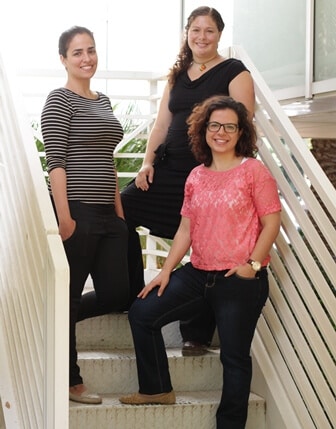
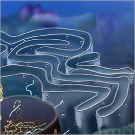
Dr. Shuldiner and Chalil Ast examined the pathways that proteins use to enter the cell organelle called the "endoplasmic reticulum": a series of folded membranes that form a labyrinthine structure where the proteins are folded, examined, and sent to their destination. The proteins passing through this organelle are eventually sent out of the cell

"The biologists hope to discover in their research 'textbook examples', meaning typical, general cases," says Dr. Maya Shuldiner from the Department of Molecular Genetics. To demonstrate her words, she opens a biology textbook, and points to a diagram depicting a molecular pathway - a series of interactions between molecules whose purpose is to bring a certain protein, or a certain process, from state A to state B. Since many proteins tend to use one molecular pathway, such "textbook" examples can reveal important insights into how the cell works, and lay the foundations for further research and discoveries.
But behind textbook examples, claims Dr. Shuldiner, a much more complex reality is hidden: "In general, the pathways that are discovered in the early stages of research in any field are considered the most important. When we discover proteins that use other pathways, we tend to treat them as outliers. Scientists often do not stop to ask, how many proteins are really used in this or that pathway".
Dr. Schuldiner and research student Tslilim Est decided that it was time to stop and ask this question. The reason for this, among other things, is the existence of new technology that allows them to test a large amount of proteins at once: the advanced microscopic equipment and the computational methods used by Dr. Shuldiner, allow her to discover the pathways used by hundreds of proteins in the cell, in a fraction of the time previously required to test a protein or two.
Dr. Shuldiner and Chalil Ast examined the pathways that proteins use to enter the cell organelle called the "endoplasmic reticulum": a series of folded membranes that form a labyrinthine structure where the proteins are folded, examined, and sent to their destination. The proteins that pass through this organelle are eventually sent outside the cell: some are hormones and external signaling molecules, and some are proteins that stop at the outer surface of the cell membrane and remain attached to it - like receptors. It is these proteins that allow the cell to sense and respond to the environment, as well as communicate with other cells. In fact, almost every human disease involves secreted proteins. The pathway by which proteins enter the endoplasmic reticulum, which was discovered in the 70s of the 20th century, is called SRP, and it has been extensively studied. Other pathways discovered since then are considered unimportant, and are simply called "SRP-independent pathways".

Is the SRP pathway indeed the main entry pathway into the endoplasmic reticulum? The scientists tested all the proteins found in the endoplasmic reticulum of a baker's yeast cell - about 1,300 proteins. The answer was clear: only about half of them used the SRP route to get there. The rest used other routes: some were identified in the past and were known, but the findings also indicated additional routes, which are not known. The scientists discovered that, at least for yeast, there is a clear division: the proteins that use the SRP pathway are the ones that remain attached to the cell membrane afterwards, while proteins that do not use the SRP pathway are secreted out of the cell. Since these pathways have been well preserved during evolution, Dr. Schuldiner believes that a similar division also exists in human cells. This means that the proteins that use the other pathways include important proteins that are secreted outside the cell, such as hormones and signaling molecules.
These days the team is trying to create a more complete picture of the alternative routes. The final goal is to identify all the molecular pathways used by all the proteins that go outside the cell. The scientists expect that the result of this research will not be a simple and ideal model, but a complex and complicated picture, which will more accurately reflect the behavior of the proteins.
This may be bad news for textbook writers, but good news for our ability to understand living cells. "Ultimately, we want to get a completely new picture of how the cell works," says Dr. Schuldiner. "We want researchers to stop looking under the torch of the accepted models, and expand the range to include all the possibilities."
traffic laws
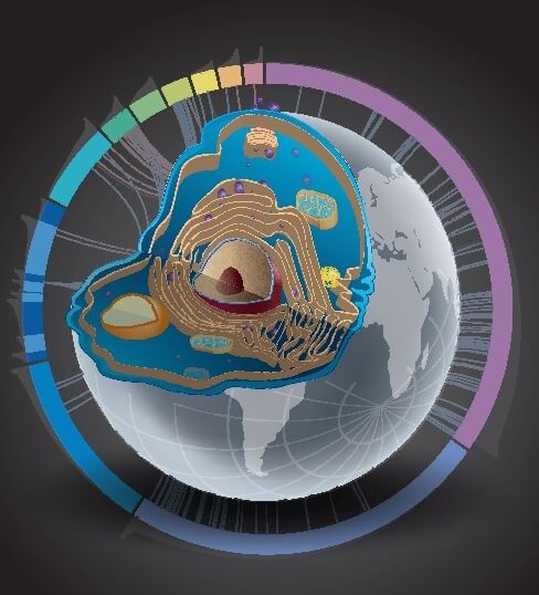
Where exactly do the proteins that circulate around the cell go? Scientists interested in this question will now be able to search for the answer. Dr. Maya Schuldiner and research student Michal Barker recently created a useful "atlas" that shows the changes in the location of proteins and their distribution in yeast cells under stress. The new atlas - which will be accessible online - presents an enormous wealth of new information, and will be an important research tool for many scientists who use yeast cells as a model for the functioning of the living cell.
Dr. Shuldiner and Michal Barker started their work with a special type of libraries - strain libraries. Libraries of this type contain strains of yeast in which certain genetic changes have been made. Baker's yeast cells contain 6,000 genes, each of which codes for making a protein. In each of the "volumes" in the strain library, a "bookmark" was placed - a genetic change was made in one of the genes in the yeast cell, which connects the protein produced from it to a fluorescent marker, which glows with green light under a special microscope. In Dr. Shuldiner's laboratory, which is equipped with advanced automatic microscopic equipment, it is possible to examine all the yeast strains in the library at once. There are other types of libraries, including those where one of the genes is deleted in each "volume". Mixing and combining different libraries makes it possible to create new genetic compositions. The entire array is then automatically scanned by a robot to discover which proteins are produced in any given experimental condition and where they are located in the cell.

This unique setup allowed Dr. Schuldiner and Michal Barker to follow the movement of proteins - one of the biggest questions in cell biology. According to Dr. Shuldiner, accurate information about the movement and location of proteins in the cell can help answer several important research questions: How many of the cell's proteins are mobile? Under what conditions do they move? How does the cell use the movement of proteins to maintain its health, and continue to divide in a variety of situations and conditions?
By exposing the yeast cell libraries to different conditions and transferring them through the robotic system, the researchers were able to follow the movement of each of the proteins in the yeast cells. The result: a complete and detailed map describing the movement paths of the proteins, as well as documentation of the amounts of the different proteins formed in different situations.
An overview of the data reveals a picture of constant turmoil in the cells: at any given time, hundreds of proteins are in motion. However, the data obtained in the experiment regarding the amounts of proteins were surprising: in many of the studies conducted today, the levels of proteins are determined through experiments that actually check the amount of messenger RNA (the molecule that carries the final instructions to create a protein) - a move similar to the use in building plans, instead of in the real buildings, to map a city. However, programs may not be executed, or, alternatively, one program may be reused. Tracking the proteins themselves revealed to scientists that the amounts of messenger RNA and the amounts of proteins do not always correspond to each other. Dr. Shuldiner: "Many studies have already shown in the past that the production of proteins is controlled in many ways even after the stage in which the RNA molecule leaves the cell nucleus. Our new findings imply that the last control steps may be more important than previously thought in determining the level of proteins in the cell."
The online atlas is called Loqate LOcalization and Quantization Atlas of the yeast proteome. "The atlas can be used by scientists who wish to investigate specific questions, such as: Which proteins are involved in a certain cellular activity? When and where do they work?" says Dr. Schuldiner. "In addition, scientists who wish to get a clearer picture of life in a cell by combining different types of information will find it an essential tool."
Power calculation
Contemporary research methods in the field of molecular biology have advanced it beyond the first step of scientific research: raising a hypothesis and testing it through an experiment. The use of fast, powerful, and fully automatic equipment, most of which was built according to Dr. Shuldiner's special needs, allowed her and the members of her group to examine all the possibilities at the same time, and to extract significant information from them. "Thanks to these tools, the research is completely freed from bias," she says. "If we once started the work with an educated guess, for example, with the hypothesis that protein A affects protein B, and then tried to prove the hypothesis through an experiment, now we can ask 'which proteins are involved in the activity of B?' Thanks to this we may discover that proteins C, D and E also affect protein A. If in the past a research student devoted his entire research work to testing a narrow hypothesis, today he can receive answers to broad questions within a few weeks."

3 תגובות
Even if there is such a camera (which is doubtful for reasons of quantum physics and the uncertainty principle), there will still be a need for someone to separate the tens of thousands of molecules, map where and where they "travel", draw atlases for the benefit of other researchers, sort out the subsystems in the cell to which the interactions belong the difference, you will interpret the general meaning of the action, you will find problems in the system that cause diseases, you will initiate the search for suitable molecules to treat those problems, you will test the hypotheses of how these molecules work (which can now be called by the explicit name "drug"), you will issue a patent for the molecule, you will issue a regulatory approval to use the medicine (by the FDA and the guys) and...come on, upload the medicine to the internet and allow the universal assembler to create the medicine for all those in need.
In short, new technology is nice, but it needs someone to operate it.
I wonder what all this will be worth in the future when they have a camera that is able to photograph and record everything that happens inside the cell down to an atomic level.
It amazes me every time how little we know about what is happening inside us...