Advanced MRI * A new diagnostic method developed at the Weizmann Institute and approved by the FDA, allows not only to determine if there is a tumor, but also if it is benign or malignant
Anat Or

A new method for identifying and diagnosing tumors developed at the Weizmann Institute has received FDA approval and is beginning to be used in breast and prostate cancer tests. The method may change the procedure of sending for a biopsy and thus save many resources, invasive tests and stress experienced by the subjects until they receive the result.
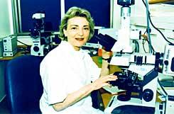
The method, which was developed by a research group headed by Prof. Hadassah Degani in the Department of Biological Control at the Weizmann Institute, is based on magnetic resonance imaging (MRI) and produces far more advanced results than those known.
The form of the MRI test is the reflection of radio waves from the hydrogen atoms in the water molecules in the body using a powerful magnetic field and receiving visual imaging.
The same equipment is also used for an MRS test, in which a radioactive contrast material is used that is injected into the subject, penetrates the spaces between the cells and enables a clearer identification of tissues with an especially high cell density - and thus the identification of cancerous tumors.
The group led by Prof. Degani used the existing MRS technology, but the form of the test changed. The existing method could detect whether there was a tumor or not, but could not tell whether it was benign or malignant. In the case of breast cancer, samples from all the tumors were sent for biopsy and nerve-wracking days passed for the test subjects waiting for the result that could change their lives.
The group of researchers discovered that the speed and manner in which the substance enters and exits the body can teach a lot about the nature of the tumor. Prof. Degani explains that malignant tumors are characterized by the fact that they are not uniform; A benign tumor, on the other hand, will be stained with the contrast material very quickly in a uniform color. When an area suspected of being a tumor is observed where the material spreads unevenly, it may be cancer. The group discovered that the exit of the material also differs between a benign and a malignant tumor; The flow of the material indicates the properties of the tissue. In healthy tissue, the substance will hardly leak out, while in a cancerous tumor, the density of the cells will lead to its rapid leakage. Since the contrast material travels through the blood vessels, the culture of microscopic blood vessels around the tumor, which characterizes cancerous tumors, can also be seen.
Besides the innovation of tracking the path of the contrast agent, the researchers also devised a special sample form that gave the test its name. The method is called 3TP (3 TIME POINTS). To get more detailed results and increase the resolution level of the image, they sample at three predetermined time points and thus allow the device to invest more power in providing a shorter but much sharper image. Decoding is done with the help of mathematical formulas.
In the clinical trials conducted in recent years comparing the results of the method with biopsy results conducted on the same subjects, the test was able to identify almost 100% of the cancerous tumors that are five millimeters or more in diameter. In experiments that also included patients with DCIS, which is cancer of the milk ducts in the breast, which does not spread and does not produce lumps - the accuracy rate was about 90%.
Prof. Degani dealt mainly with breast cancer and the new method has many advantages over the existing methods for diagnosing the common disease. Degani believes that in the future the magnetic resonance and ultrasound methods will replace the X-ray, which is currently the cheapest method but has the risk of ionizing radiation.
Of the samples sent for biopsy, 80-65% are of benign tumors. The new test will be able to save the invasive procedure and the wait for many of the women in whom a tumor is found. Degani mentions that in a small part of the situations the answer will be borderline and it will not be possible to determine with certainty the type of tumor using the 3TP method and then a sample will be sent for biopsy. "But there will be a fairly large group of tumors that we can say are benign."
After the end of the research on breast cancer, which lasted more than 10 years, the team turned to testing prostate cancer, which in terms of the nature of the tissue and tumors is not very different. Research is now starting on lung cancer, which is the deadliest cancer in the world and the detection of which is often late. "It's a difficult subject," says Degani, "the tissue itself is not an obstacle, but the strong movement of the breath and the heart creates a technical problem." The tests with the new method came into use in the United States but not yet in Israel.
