Magnetic resonance research at the Weizmann Institute has been at the forefront of this field for decades. These days, four new magnetic resonance devices are being installed in the institute, one of the most powerful in the world today, and with their help unprecedented clinical and biological research will be possible. "However," says Prof. Lucio Friedman From the Department of Chemical and Biological Physics, "Even when we study certain tissues and organs using MRI, even in the most powerful systems, we face significant challenges, and we work vigorously to overcome them."
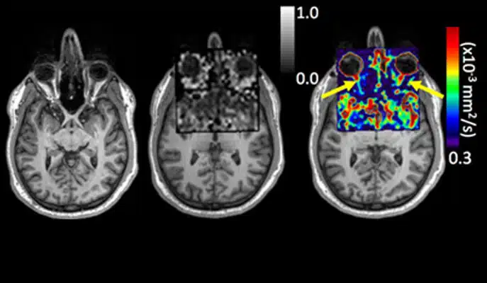
It should be known that the MRI device works optimally when it comes to the imaging of homogeneous tissues; That is, tissues where water molecules and metabolites are uniformly distributed in the magnetic field. However, not all parts of the body are homogeneous, and some even sometimes include air spaces; Such are, for example, the nasal passages and facial cavities, the areas of the base of the brain, the areas near the optic nerves and the olfactory bulb, and most of the abdominal cavity. Usually, MRI systems fail to overcome the distortions in the magnetic field resulting from the lack of homogeneity, and this problem even worsens as the intensity of the magnetic field increases. Because of this, the power of the modern equipment does not have any advantage in this sense. These limitations are particularly noticeable in scanning techniques that rely not only on the location of the molecules, but also on mapping their diffusion, that is, mapping the extent and direction of water movement in the tissues. Diffusion analyzes are of great importance, because they can teach us about the structure of the brain, and indicate the presence of small tumors or the development of nerve cells. But, as mentioned, in non-homogeneous tissues, such as those in the brain, the imaging will often show distorted data.

Prof. Friedman and his team have been working for a decade on finding a solution to this problem, among other things, using the xSPEN method - short for cross-term SPatiotemporal ENcoding - which makes it possible to produce reliable simulations even in non-homogeneous tissues. To create these simulations, adjustments and changes must be introduced in all the critical stages of an MRI scan: from the pre-coding stage of the information before the scan, through the creation of adapted patterns of the changes in the intensity of the fields in the scan, and ending with a new data processing algorithm for converting the MRI signals into images. "The development of these new techniques involved quite a bit of programming," says Prof. Friedman, "but first and foremost it relied on breakthroughs in physical understanding, which brought up alternative ways of imaging through magnetic resonance. This is how a new approach to MRI was born, thanks to which it is possible to obtain a more accurate imaging: for the first time, instead of trying to overcome the problem by force or trying to circumvent it with corrections after the scan, the method provides a solution by including the inhomogeneity in the imaging process itself."
The results of the method demonstration, which were published recently in the scientific journal Scientific Reports, clearly illustrate xSPEN's ability to create images of particularly challenging areas of the human body, such as the peripheral nervous system and the head, including the fine optic nerve leading from the eye to the brain. Prof. Friedman's research group works to adapt the technology to the imaging of organs and other inhomogeneous areas, including the placenta, which serves as a mechanism for transferring nutrients from the mother to the fetus.

Prof. Friedman and his team are collaborating in this research with two hospitals in Israel with the aim of turning all the stages of the method into one simple process that is easy to operate, so that technicians in the hospitals can use it after only minimal training. And here, Prof. Friedman explains, the new method has a great advantage, because it is completely compatible with the existing hardware.
Ten times more sensitive
The institute's magnetic resonance activities - electron electromagnetic resonance (EPR), nuclear magnetic resonance (NMR), and magnetic resonance imaging (MRI) - are currently in the midst of upgrades, which include the installation of an innovative MRI scanner with a 15.2 tesla magnet for animal research, powerful scanners of 3 Tesla and 7 Tesla for human use, and a 23.5 Tesla (1 GHz) system for NMR research.

Prof. Friedman is very optimistic about the future of MRI research: "In recent experiments, for example, we have seen that the new 15.2 Tesla system improves the sensitivity tenfold, compared to previous results with the 7 Tesla system, and the greater sensitivity will allow better spatial separation (resolution)." Different research groups at the institute plan to use the new systems in a variety of projects - from developing new biosensors for the early detection of diseases, to monitoring the formation of new blood vessels or monitoring brain activity.
#Science_Numbers
A 7 tesla MRI machine for human use produces a magnetic field 140,000 times stronger than the Earth's magnetic field.

One response
I would be happy to know where this mri can be done because I had brain surgery in the area of the optic nerve and the entire tumor was not removed