Collaboration between universities in the US resulted in the creation of new hybrid nanosubmarines capable of identifying and killing cancer cells, marking them with iron grains for MRI examination and phosphoric molecules for surgery

You wake up one morning and go to the doctor for your annual routine checkup. He checks your weight, pulse, blood pressure, blood cholesterol level and finally injects you with a small ampoule, filled with billions of tiny nano-submarines. The nano-submarines are dispersed in the bloodstream and probe for tumors and cancer cells. When they find such, they cling to them, penetrate them and release a deadly poison inside the cell.
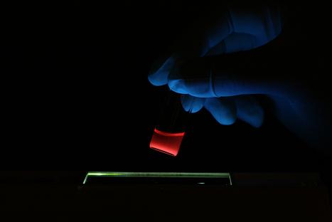
To verify the success of the treatment, the doctor takes you through a fast MRI. In addition to the poison they contain, the nano-submarines also carry paramagnetic iron grains that appear on an MRI and indicate their location in the body, where they have bound to the cancer cells. The MRI shows several tumors that were too large to be destroyed by the nanosubmarines alone. There is no choice: the tumor must be analyzed and removed from the body. But even this operation, which usually requires an expert oncologist, becomes extremely simple thanks to the nanosubmarines. Each of the tiny submarines also contains phosphorous molecules, in addition to the poison and the iron grains. The doctor uses special glasses that allow him to see the tumor glowing orange inside the body, and removes or burns it with a laser without leaving even a single living cancer cell in the body.
According to all the advances in the field of nanomedicine, there is no doubt that the described future is approaching us with giant steps. Different types of nanosubmarines have already been described in the past, including iron 'nanoworms' that identify tumors and mark them for the MRI examination, nanosubmarines containing a poison that kills tumors and even nanosubmarines that glow in the dark. Now, in a combined study from the University of California San Diego, Santa Barbara and MIT, all these features have been combined together to create a nano-submarine capable of doing everything at once: stick to a tumor, secrete a toxin into its environment, mark it for detection by MRI and remove it from the body by fluorescence (phosphorus).

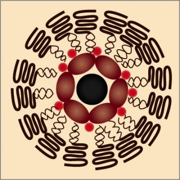
"The idea combines imaging agents and drugs into a protective 'mother ship' that avoids the natural processes [in the body] that would eliminate these agents if they were unprotected," says Michael Saylor, professor of chemistry and biochemistry at UC San Diego. Saylor headed the team of chemists, biologists and engineers who turned the extraordinary idea into reality. "The diameter of the mother vessels is only 50 nanometers - 1,000 times smaller than the diameter of a human hair. They are equipped with an array of molecules on their surface, which allows them to discover and penetrate tumor cells in the body."
The 'shell' of the nano-submarines consists of lipids - fatty molecules that make up the surface of all body cells. The lipids in this study were specially modified, allowing the submarines to drift in the bloodstream for many hours before being eliminated, as demonstrated in a series of experiments on mice. The ability of the submarines to evade the body's immune systems is particularly important, because it allows them to import drugs in high concentrations and over time to the tumor cells.
"Many drugs look promising in the lab, but fail in humans because they don't reach the diseased tissue in time, or in high enough concentrations," explains Sangeeta Bhatia, a physician, bioengineer, and professor of health sciences and technology at MIT, who played a key role in the development of the nanosubmarines in his lab. . "These drugs do not have the ability to evade the body's natural defenses or differentiate between the target cells and the healthy tissues. In addition, we lack the tools to diagnose diseases, such as cancer, in their earliest stages of development, when the drugs can be particularly effective."
To optimize drug delivery to cancer cells only, a protein called F3 is attached to the surface of the submarines. This protein, engineered in the laboratory of Arki Ruoslati, recognizes the cancer cells, binds to them and is transported into their nucleus together with the submarine. This protein is only the first generation of proteins that identify tumors and enable their effective treatment.
"We are now assembling the next generation of nanodevices for smart tumor detection," says Ruoslatti, a professor and biologist at the Burnham Center for Medical Research at UC Santa Barbara. "We hope that these devices will improve the diagnostic imaging of cancer and allow precise targeting of treatments for cancerous tumors."
The submarines were loaded with three different types of cargo before being injected into the mice. Two of the payloads are nanoparticles of different types: superparamagnetic iron-oxide grains, and phosphorescent quantum dots. The third cargo is doxorubicin, an anti-cancer drug. The iron nanoparticles enable detection of the submarines in an MRI, while the quantum dots can be detected using a fluorescent scanner.
"The fluorescent image provides a higher resolution than the MRI," Saylor says. "One can imagine the surgeon checking the exact location of the tumor in the body before surgery using MRI, then using fluorescent imaging to find and remove all parts of the tumor during surgery."
The members of the research group discovered to their surprise that a single mother ship can carry several grains of iron oxide, which increase its brightness in the MRI image. Despite the fact that there are already hybrid nanosystems capable of carrying different types of nanoparticles, there are very few experiments done with hybrid nanosubmarines on living organisms.
"This study provides the first example of a single nanometer material capable of delivering drugs and enabling multi-mode imaging of diseased tissue in an animal," says Ji-Ho Park, a doctoral student in Saylor's lab who participated in the study. The researchers are now working on coating the nanosubmarines with chemical recognition molecules, which will allow them to reach specific tumors, tissues and organs in the body.
For information on the website of the University of California in San Diego
On the science website:

9 תגובות
point,
Are you infected from Hogin?
But if we are healthy, what is the point in cellular submarines?
Someone has to be sick even if they are decent, and it is not fair that only we will be healthy.
Dear Ami Bachar
As for BBB, oops, I suddenly realized how to switch to English in the blink of an eye (not the printing type)..
I wondered which one of us is the pure scientist, you, who hides behind nanotube formulas, and numbers. Or maybe I'm really selfish after all, yuck.. the one who researches in the capillaries of your being and doesn't allow you to escape from pure research..
If I'm wrong, please correct me.
The main thing is that we are healthy.
Hugin
Ami,
To look into such a submarine a penetrating electron microscope is needed. Most likely such a hybrid submarine will contain the above components more or less as shown in the picture, but of course with more mess.
About the MRI, you are right. The hope is that if it is such a small tumor (100 microns is less than ten human cells sitting one after the other), then the submarines will be able to kill it themselves. As for the BBB, this is indeed a sore point in the field of nanosubmarines. But let's tell the truth, if only the cancerous tumors in the brain make up the totality of cancer morbidity in the 21st century, we will discuss it.
Just let's be healthy.
It is interesting and nice to hear that there are beautiful advances in the field of nanotechnology of man and his health.
We recently heard about microscopes with very high capabilities to separate with a resolution of a single angstrom and even less. My question is: when you look at such a submarine with a special microscope that is able to see it in detail - is the structure shown here as a model close or similar to what you find in the test tube or on the slide?
First of all, I will say that the resolution of MRI, as far as I know, is around a hundred microns, so when it comes to a very small cancerous area, it may not be possible to pick it up at all. Moreover, the entire area of the brain and the barrier (BBB) is also a problem. Good luck and stay healthy.
Ami Bachar
Although liposomes have been in the field for a long time, but as you mentioned, here it is a much more complex technology with many more options.
5-10 years is a very short time...
We'll wait and see…
Hanan
Hanan,
The technology of the nano-submarines is already used today in medicines for humans. The most primitive nanosubmarines are liposomes, which have been used in medicine for over 30 years. All of the above is just an improvement and streamlining of this technique.
I personally have not heard of clinical trials on humans with the most innovative nanosubmarines, but I assume that there are already some, and that in 5-10 years we will get to start reaping the fruits of the current efforts in the laboratories.
Shame on those who die the day before the launch.
Very interesting. If the technology is implemented and developed, it foresees fundamental changes in the treatment of diseases.
Are there prospects for clinical trials in humans?
Hanan
http://WWW.EURA.ORG.IL