A journey through the human body is no longer a fictional dream. It is possible that tiny devices will perform surgeries, deliver drugs, and help diagnose diseases in the near future

By Paolo Dario and Ariana Manciasi
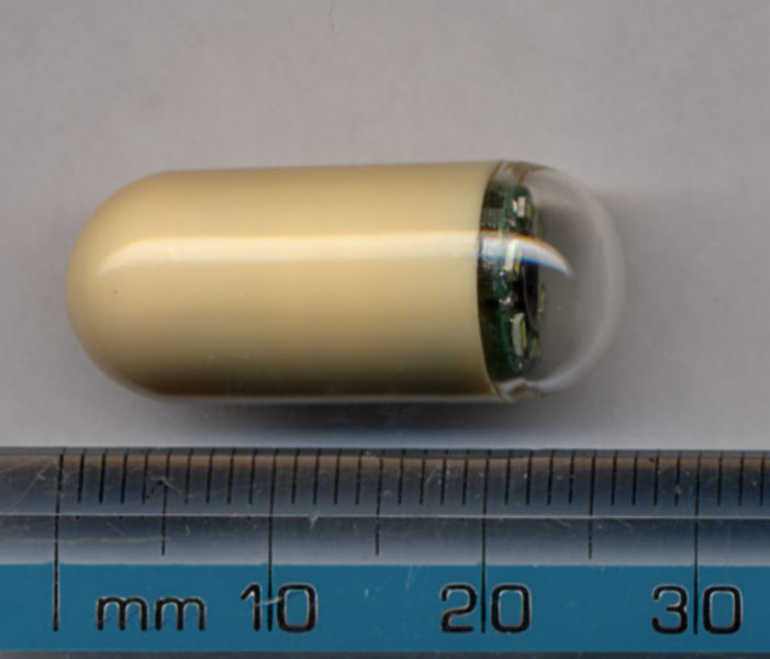
The movie "Amazing Journey," which depicted a team of miniature doctors sailing through a patient's blood vessels to make life-saving repairs to his brain, was pure science fiction when it was released in 1966. In 1987, when Hollywood made the movie "A Day in the Trap", based on a similar idea, real engineers had already started building prototypes of pill-sized robots that could travel through the patient's digestive tract to assist the doctor. Patients began swallowing the first commercial camera pills in 2000, and since then doctors have been using such capsules to see places that had not been seen before, the inside of the small intestine for example, places that are very difficult to reach without surgery.
But one important aspect of "Wonderful Journey" still remained fictional at the time: the idea that these tiny pill cameras could move on their own, swim to a tumor to take a biopsy, determine the nature of inflammation of the small intestine and even treat an ulcer medicinally. However, in recent years, researchers have made great progress in converting the basic principles of the passive pill camera and in the development of active tiny robots. Today there are advanced prototypes with legs, propellers, sophisticated photographic lenses, and wireless guidance systems and they are already being used for experiments on animals. Soon, it is hoped, these tiny robots will be ready for clinical trials. Currently, scientists are pushing the boundaries of tiny robotics.
The transformation of the passive pills
The digestive system is the first front. The first wireless pill camera, M2A, manufactured by the Israeli company Given Imaging, and later models also proved the effectiveness of stomach and intestinal examinations using a wireless device. The process, called capsule endoscopy, is already a matter of routine. Unfortunately, since there is no control over the pill camera, there is a high rate of false negative results. The cameras may overlook problematic areas, and are therefore not a reliable diagnostic tool. If the doctor asks to look inside the body to scan and look for diseases or to look closely at a suspicious place, the most important ability for him is the ability to stop the camera and direct it to the area of interest.
Turning the passive pill into a device that can provide more thorough scans of the digestive system requires the addition of moving parts, mechanisms that will move the pill in the body or be used as tools for tissue treatment. Operating these moving parts requires wireless, fast, two-way transmission of data, instructions, and images. The pills should basically be tiny robots capable of responding quickly to the operator's instructions. All these components need a source of power sufficient to carry out the task assigned to them to the end, in a journey that can take up to 12 hours. All of this needs to fit into a two milliliter (ml) capsule (about the size of a gummy candy) so that the patient can swallow the device relatively comfortably.
In the year that M2A was put into use, the "Center for Smart Micro-Systems" (IMC) in Seoul started a ten-year project to develop a new generation of advanced endoscopic pills. For the purpose of photography, such a robotic pellet will be equipped with sensors and a light fixture. It will include mechanisms for administering drugs and collecting samples. It will be able to move independently under the wireless control of the test operator. Since 2000, more companies and research groups have entered the field. Eighteen teams from Europe, for example, established a consortium with the IMC to develop robots in a pill to detect and treat cancer. Our group, from the Higher School of Sainte-Anne in Pisa, Italy, under the supervision and medical direction of Mark A. Schur from Tubingen, Germany, technically and scientifically coordinates this project, called VECTOR (a multipurpose endoscopic capsule for the detection and treatment of tumors in the digestive system.)
These teams, from academia and industry, came up with many innovative ideas. And in particular, they proposed a variety of solutions to the main challenge: how to control the movement of the pill device that wanders through the body's recesses. Basically, most take one of two approaches.
The first approach involves moving the capsule through mechanisms installed in it: moving parts such as paddles, legs, propellers, or similar accessories that are integrated into the device's shell and can come into action after the pill has entered the digestive tract. The mechanisms driven by tiny motors are usually used to direct the movements of the capsule, but there are models in which the legs can also make a way for it in the surrounding tissue, allowing it to approach certain places or pass through a section where the intestine has collapsed and blocked. The total volume of an ingestible pill makes it impossible to insert motors and mechanisms such as relays, therefore the installation of other necessary parts, such as photo sensors or a medical component (such as a device for taking a biopsy), is not an easy challenge. Also, to expand tissues, the capsule needs to exert a considerable force: 10 to 20 times its weight. Only motors with a large torque can produce such power, and these consume a lot of energy (about 0.5 watts). This energy consumption burdens the battery technology, and the duration of the device's operation is shortened.
To save energy, the best compromise is, perhaps, to use mechanisms only for propulsion, and to do tissue stretching in another way. If the patient is allowed to drink half a liter of clear liquid, before swallowing a capsule driven by a propeller, for example, the stomach will expand for 20 minutes, or a little less, until the liquid drains into the small intestine. During this period of time, the pill will be able to swim to her, with wireless guidance, and examine the stomach wall.
The problem of the size of the built-in mechanisms and their energy consumption led some of the researchers to base themselves on a different approach: using a magnetic field that originates outside the body to control the movement of the capsule. The companies Olympus and Siemens presented for the first time in 2005 a magnetic guidance system for a passive camera pill that they produced. According to Siemens publications, the system rotates the pill on an axis. This screwing movement creates a slight friction that helps the capsule to enter narrow sections of the digestive tract, for example the small intestine.
Although the use of magnets to guide an endoscopic capsule in intestinal papules is quite simple, it is very difficult to achieve good control using magnets alone. Magnetic fields weaken as you move away from their source, and due to the crooked and tortuous route of the intestine, sudden changes in the strength of the field can bounce the capsule, and the effect of the field can even be canceled completely. In practice, such instability may result in the connection between the operator and the capsule being severed and it being lost forever. This can be compensated for by adding magnets to increase control and stability, but this requires cumbersome arrays of electromagnetic coils.
Hybrids according to size
In view of the limitations of the two approaches to driving the capsule, i.e. the control from the inside and the control from the outside, we believe that they should be combined to create a device that is comfortable to swallow and provides reliable diagnoses. Movement using external magnets is good for rough positioning of the capsule in the intestine. Leg-like mechanisms are good for changing position and maneuvering to improve the image.
Our research group therefore designed such a hybrid capsule, with four motor-driven legs. We tested it in a pig, an animal whose intestines resemble a human intestine. The legs of the capsule are folded during swallowing and during most of its journey through the digestive tract. An external magnetic field generator is attached to the stomach and guides the capsule forward. When the capsule reaches the place where the walls of the intestine have collapsed, it spreads its legs and lifts the tissues around it, and they also push it forward a little through the created opening.
In most areas of the small and large intestine, such a mixed drive system can provide the necessary control for doctors to visually inspect. Every problem has its own solution. In the VECTOR project, for example, they developed three different ideas just for the capsules intended for the small intestine: one is a passive camera pill for normal scanning, the other is a diagnostic capsule with an active drive and a spectroscopic camera that can detect abnormalities below the surface of the tissue. The spectroscopic camera is also integrated in the development of the third concept of the VECTOR capsule. This device will also be equipped with a biopsy accessory, which can take a sample of tissue and store it inside the capsule until it leaves the patient's body.
If the endoscopic pill robots have the ability to perform biopsies and other complex healing operations, such as surgical procedures, they will be an even more useful and efficient tool in the hands of doctors. But due to crucial problems such as the energy supply, volume constraints and too weak a torque, it is impossible to perform more ambitious healing operations in one pill with a volume of two ml, which require complicated movement and multiple mechanisms.
That is why we are currently working on a more advanced solution: surgical robots that change their configuration inside the body. It will be done, perhaps, like this: the patient will drink a liquid to expand the stomach, then swallow 10 or 15 pills. Inside each pill there will be a tiny component and at both ends there will be two magnets. When the capsules arrive in the stomach, they will immediately join each other, through remote control, and assemble the desired configuration. The surgeon will therefore be able to perform an operation using the assembled robot, without having to cut the body from the outside at all. After the surgery, it will be possible to change the configuration of the magnetic connections between the components, or to repair and let the parts leave the digestive system without causing any damage.
We have an initial prototype based on capsules that are two centimeters in diameter, with internal parts and mechanisms that can be adjusted as needed. It is possible that one of the pods, or more, will carry a camera, others will carry other tools and all will be wirelessly controlled.
Tiny robotic components could eventually be used for a wider range of actions, throughout the body and for all kinds of purposes. Guidance systems and camera sensors developed for pill endoscopy are already influencing medical technology in adjacent fields, such as, for example, in the newest version of the endoscope and the laparoscopic surgery tools. Outside of the medical world, these technologies are part of a broader trend toward miniaturization and wireless control of multipurpose robots. There is no doubt that the robots in the pill will have an impact on the world of robotics in general.
__________________________________________________________________________________________________________________________________________________________________________
key concepts
Camera pills make it possible to see the entire digestive system from the inside, an unprecedented possibility, but the uses and accuracy of the passive capsules are limited.
Today, active robotic capsules are being developed, the size of a pill, capable of scanning, diagnosing and performing medical operations.
The miniaturization of robotic components that will fulfill tasks inside the body involves overcoming new engineering challenges. These challenges give rise to creative solutions that will affect robotics and other medical technologies in general.
At work
Tiny robots for a wide variety of tasks
To make tiny robots that can operate in the digestive system, engineers must find ways to wirelessly control their movements. They also need to insert suitable tools, photo sensors and an energy source into an ingestible capsule. Here are some examples of the varied tasks that the engineers want to assign to the tiny robots and the ways in which they try to overcome the technical difficulties.
movement
The movements of the endoscopic robot can be controlled by mechanisms installed in the device itself, such as legs, paddles, propellers and eyelash-like devices, or externally, using magnetic fields that originate outside the patient's body.
tissue expansion
One way to move aside tissue and level a path to improve the perspective is to equip the robot with strong arms that can push. A more energy-efficient method is to have the patient drink water to expand the digestive tract enough to maneuver the propeller-driven capsule.
diagnosis and treatment
A capsule can carry various devices: a spectroscopic camera, which sees cells under the top layer of the tissue, scissors for taking tissue samples, or a container containing medicines.
About the authors
Paolo Dario and Ariana Menciassi are professors of biomedical robotics at the Sant'Anna College in Pisa, Italy. Dario, who invented the first self-propelled colonoscopy robot in 1990, was also a pioneer in the development of robotic capsule endoscopes in his work with the "Center for Intelligent Microsystems" of South Korea and in partnerships with other European robotics researchers. Manciasi, who has been working with Dario for 10 years, specializes in micro-engineering for minimally invasive treatments and medical nanotechnology.
remote analysis
One of the ways to increase the variety of tasks the pill robots can perform is to design them to assemble themselves. The patient will swallow about a dozen pills or more, and when they reach the stomach, they will join each other into one big and strong robot. The surgeons will operate the device wirelessly. At the end of the operation, the robot will return and break down into separate capsules, and these will leave the digestive tract without causing any damage.
More on:
Wireless Capsule Endoscopy: From Diagnostic Devices to Multipurpose Robotic Systems. Andrea Moglia, Arianna Menciassi, Marc Oliver Schurr and Paolo Dario in Biomedical Microdevices, Vol. 9, no. 2, pages 235–243; December 12, 2006.
Wireless Therapeutic Endoscopic Capsule: In Vivo Experiment. P. Valdastri et al. in Endoscopy 2008, Vol. 40, no. 12, pages 979–982; December 2008.
Wireless Reconfigurable Modules for Robotic Endoluminal Surgery. Kanako Harada, Ekawahyu Susilo, Arianna Menciassi and Paolo Dario in Proceedings of the 2009 IEEE International Conference on Robotics and Automation. Kobe, Japan, May 2009.
