A pair of researchers at MIT have developed a new system that uses a focused laser beam to suspend and sort cells. The system enables new biological studies, and provides an inexpensive method for optimizing clinical tests and diagnostics, genetic screening and clone research
How do you find a needle in a haystack?
A similar problem is often faced by biology researchers, who have to examine millions of different cells, find the single cell that is different from all the others, and separate it from the others. Now a new sorting method has been invented at the Massachusetts Institute of Technology, which uses a laser beam to separate the cells.
The developers – Joel Waldman, a professor of electrical engineering and computer science at MIT, and Joseph Kovac, a graduate student in the same department – claim that the system can sort up to 10,000 cells on a standard microscope slide, opening the door for new biological studies. , which were not possible until now. The possible applications are in clinical tests and diagnostics, genetic screening and clone research. All these fields require the selection of cells with certain properties. The system is described in a main article in the journal Analytical Chemistry published on December 15.
In the methods used today, a fluorescent marker (glow) is used that identifies certain proteins that appear only on the cells that you want to mark. The new system enables more accurate sorting, by separating the cells not only based on the level of light they emit, but also based on the various activities that occur in specific areas of the cell, such as the nucleus. The system can also detect the reaction speed of cells, or how long the reactions last.
"We wanted to look at things inside the cell that change over time, or that are in certain places," says Waldman. With today's technologies it is not possible to separate cells according to these features.
The new system allows, for example, to separate cells according to the speed of their reaction to a certain fluorescent substance. "The system will be able to identify the cells that respond quickly or slowly, and measure the difference," says Waldman. "It sounds like a simple task, but it really isn't." There are other ways to separate cells in a similar way, but they require complicated and expensive equipment, or are limited by the number of cells they can work on.
The new system uses a transparent silicone layer attached to a standard microscope glass. Many tiny holes were designed inside the silicon layer, which are actually small traps in which the cells are trapped when they are transferred to the glass from the solution. Each cell finds another small hole for itself and is trapped inside. Up to 10,000 cells can be sorted on one microscope slide.
The technician looking through the microscope can examine each cell and determine its properties. It can determine, for example, whether different parts of the cell glow as a result of the fluorescent staining, or the speed at which the cell is filled with fluorescence - that is, the rate at which the fluorescent marker adheres to different factors in the cell. A simple computer program can make the same diagnoses. The position of the various cells is marked by the computer, and at the end of the computerized marking process, the practical selection process is activated. All selected cells are floated out of their traps. The levitation is carried out by a focused laser beam, which is operated at low energy. Due to the tiny size of the cells, the concentrated light beam can push them upwards, out of the trenches in which they are trapped. From the moment the cells emerge from the holes, a liquid is injected that sweeps the suspended cells into a separate container.
Although the laser is capable of applying force to the cells, it does not have enough energy to harm them. Even after the separation, the cells on which the laser was applied remain alive and intact, and are able to divide and continue their normal biological activity.
The uses of the system are numerous. One of the main uses could be to separate different types of cells created from a single stem cell, thus streamlining the medical cloning process. Another possible use is the separation of cells detected in different blood tests. This procedure can also result in the selection of red blood cells that carry malaria parasites, thus allowing for better research of the disease. Another possibility is in the study of intracellular processes, since the system can differentiate between processes that occur in the cell body and the processes that affect the most important treasure of the cell: the DNA inside the nucleus itself, which contains the operating instructions for the entire cell. If a powerful enough microscope is used, it is quite possible that the system will be able to differentiate between cells at different stages of receiving signals from outside the cell, thereby advancing the science of molecular biology.
Waldman and Kovac continue to perfect the system, with work currently focused on ease of use and improving the sterility of the system. Waldman says that unlike other expensive separation techniques, such as optical tweezers that can only be found at large research centers, the new system can cost only a few thousand dollars. He predicts that they could be used in many biological research laboratories, and even in clinical clinics.
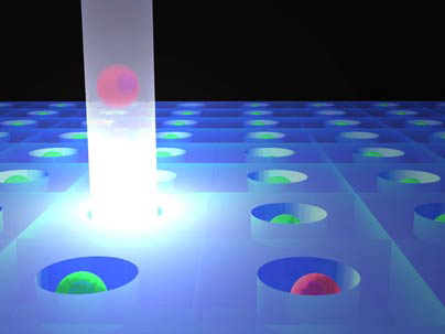
Image 1: MIT has developed a new system for cell selection, which incorporates special 'traps' in a silicon layer attached to a microscope glass. Cells with the desired properties are forced out of their pits with the help of the pressure exerted by a focused low-energy laser beam. After the cells are floated out of the holes, the silicon layer is washed with liquid that sweeps the cells into a separate container. Image courtesy of Joseph Kovac, MIT.

5 תגובות
Nobel prize?
Thanks. It's always nice to be right.
"When I finished the news, I looked at it again and told myself that this is an invention that most of the readers of 'Hidan' will not regard as something big."
Right.
Yes indeed.
When I finished the news, I looked at it again and told myself that it was an invention that most of the readers of 'Hidan' would not regard as something big. But biologists like me and you, who have to deal with fluorescent microscopes, FACS, ELIZA and the like, will see it as a godsend.
Of course, you should wait and see where things stand. I have a feeling it's going to be a while before we get to see the invention in labs outside of MIT.
Amazing and genius in its simplicity! Until today we used FACS machines to sort up to 4 cells and that too in the best case. I can think of many, many ways to take advantage of such a simple system. Those who can apply it will be very lucky in the coming year.