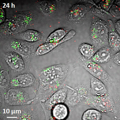
Until now, there has been no method to observe nanotubes in living cells and in the bloodstream, says Purdue University biomedical and chemical engineering professor Ji-Xin Cheng.

Researchers have demonstrated a new imaging tool for tracking materials called carbon nanotubes in living cells and the bloodstream, a development that could aid efforts to improve their use in biomedical research and clinical medicine.
These materials (carbon nanotubes) have the potential to be used in diverse applications in the fields of drug delivery to treat diseases and medical imaging for the benefit of cancer research. Two types of nanotubes are produced in the production process - metallic and semi-conducting. However, until now there has been no method to observe these two types in living cells and the circulation, says Purdue University biomedical and chemical engineering professor Ji-Xin Cheng.
The imaging method, called transient absorption, utilizes laser pulses in the near-infrared spectral range to transfer energy to the interior of the nanotubes, then use another laser pulse in the same range to examine the nanotubes. The researchers were able to overcome the main obstacles that existed in the imaging technology whose purpose was to detect and monitor the nanotubes inside living cells and laboratory mice, explains the lead researcher. "Since we are able to do this at high speed, we can see what is happening in real time as the nanotubes flow through the bloodstream," adds the researcher. The research findings were published in the scientific journal Nature Nanotechnology.
The innovative imaging method is "label free", that is - it does not require that the nanotubes be labeled with dyes, a fact that makes the method particularly suitable in research and medicine, adds the researcher. "This is an important means of research that can provide information to the scientific community, and thus it can learn how to improve the use of nanotubes for the benefit of biomedical and clinical applications," explains the researcher. The existing imaging method makes use of the luminescence mechanism and is limited since it is able to locate only the semi-conducting type nanotubes but not the metallic ones.
The nanotubes have a diameter of about one nanometer and cannot be seen with a normal light microscope. One of the challenges in using this innovative method for living systems was the need to eliminate the interference created by the background light of the red blood cells, which is brighter than the nanotubes. The researchers solved this problem by separating the signals received from the red blood cells from the signals received from the nanotubes into two distinct channels. The radiation from the red blood cells is inhibited relatively little compared to the radiation emitted from the nanotubes. The two types of signals become "phase different" by restricting them to different channels based on this delay.
"The method is important in the field of drug delivery since you are interested in knowing how long the nanotubes remain in the blood vessels after they are injected into the body," explains the lead researcher. "Therefore, you are required to locate them in real time while they are flowing in the bloodstream." The structures, called single-walled carbon nanotubes, are obtained by folding up a monoatomic layer of graphite, known as graphene. By their nature, the nanotubes are hydrophobic, and in order to use them for research, some of them were coated with DNA molecules in order to allow them to be sealed with water, a characteristic required for their transfer from the blood circulation to the inside of the cells themselves.
The researchers also obtained images of the nanotubes that accumulated in the liver and other organs in order to examine their distribution in mice, and they are taking advantage of the new imaging method to examine other nanomaterials, such as graphene itself.
