In the laboratory of Prof. Yonina Elder in the Faculty of Electrical Engineering at the Technion, a tiny and efficient innovative ultrasound system is being developed, which transmits the scans immediately to the attending physician. Such a system will make it possible to carry out ultrasound examinations in disaster areas, in the event of road accidents and in poor areas where there is no medical infrastructure, and to provide the team in the field with remote medical instructions based on the findings
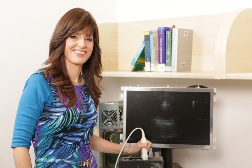
An innovative approach to ultrasound testing was developed in the laboratory of Prof. Yunina Elder in the Faculty of Electrical Engineering at the Technion. This is a sophisticated probe, which eliminates the need for the large ultrasound devices we know from clinics and hospitals. The transducer acquires only the relevant information, so that the scans received on the miniaturized device can be transmitted through the "cloud" to the treating physician's smartphone (or tablet).
Dr. Shai Yordan Taiman, a cardiologist from Sheba Hospital, explains that in the case of wounded in the field, for example, the development will provide "real-time information to a doctor who is not in the field, and will allow him to guide the paramedic who is on site. This development will make it possible to remotely treat patients in developing countries under the guidance of Israeli doctors."
Ultrasound imaging is one of the most common tests in the world of medicine. Its advantages: it is non-invasive, does not involve exposure to ionizing radiation, has no risk and is relatively low cost. The ultrasound examination is based on high-frequency sound waves that we cannot hear, hence its Hebrew name: "auditory image". During the test, a transducer that transmits sound waves is attached to the subject's body, and based on the pattern of the reflected waves, an image of the body parts being scanned is constructed. This technology is used in a wide variety of important medical tests, including assessment of the condition of the fetus in the womb, examination of the baby's brain through the iliac crest (the space between the bones of the skull), diagnosis of internal organs, assessment of blood flow, diagnosis of the thyroid gland, heart examination and detection of tumors and inflammations.
In the current procedure, the test is performed in clinics and hospitals by a transducer connected to a large, cumbersome and expensive ultrasound device. The test results are collected on a computer and deciphered by a radiologist who sends the diagnosis to the attending physician (usually the family doctor). This process takes several whole days, which may be critical in some cases.
Uploading the test results to the "cloud", through which the attending physician can view the findings from his mobile device, may save a lot of time, but so far this has been avoided due to the amount of information accumulated in each ultrasound examination. Also, the high data acquisition rate in the device requires connecting the transducer to the device with a heavy and thick cable.
The good news is that in the SAMPL laboratory, led by Prof. Yunina Alder from the Faculty of Electrical Engineering at the Technion, a system has been developed that dramatically changes the nature of the ultrasound tests. First, a new algorithm developed in the laboratory makes it possible to compress the information already at the initial scanning stage in a way that allows it to be uploaded to the cloud, without harming the quality of the image and without losing information along the way. Second, the innovative transducer developed in the laboratory eliminates the need for the large ultrasound device currently used in most clinics.
Prof. Yonina Elder's laboratory focuses on developing innovative methods for processing information using only a small part of the sampled information. Reducing the amount of sampled information has very dramatic positive consequences: shortening the duration of acquiring the information and processing it, miniaturizing the systems and speeding up their activity, reducing the required electrical power and saving money. The "wisdom" here is of course finding ways to recover the initial information even though it is not transferred in its entirety in this process. The laboratory researchers are working on this, and now, as mentioned, a dramatic achievement has been recorded in the field of ultrasound.

2 תגובות
Yunina Elder. A real cannon.
It is worthwhile to develop a device that will be in every HMO branch and perhaps in every family doctor. Will save a lot of patients running around and save the 17 form.