The discovery may contribute to the future development of advanced ways to heal muscles using stem cells

The development process of muscle tissue in the fetus consists of several stages. In the first stage, the primary embryonic cells differentiate into cells that will become muscle cells. The cells found at this stage in the differentiation process are called myoblasts. In the second stage, the myoblasts recognize each other and adhere to each other. In the third stage, fusion takes place, in which the membranes between the cells come together and burst, so that unified, large, multinucleated cells are formed. These united and united cells are the muscle fibers that are able to contract, exert force and perform work.
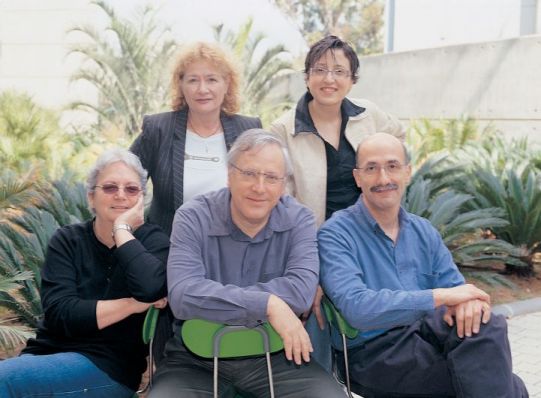
The way in which myoblast cells recognize each other and adhere to each other was known, but the way in which the membranes fuse remains a mystery. A study recently carried out by Weizmann Institute of Science scientists sheds new light on this process. Research student Rada Masarva and laboratory technician Shari Carmon participated in the study, under the direction of Dr. Eyal Schechter and Prof. Ben-Zion Sheila, from the Department of Molecular Genetics at the Weizmann Institute of Science. Dr. Vera Shinder from the Electron Microscopy Unit also participated in the study. The mutual recognition process of the myoblast cells is based on protein recognition molecules found in the cell membranes so that one end of them sticks out, while the other end is inside the cell. When two such recognition molecules adhere to each other, the cells anchor side by side, in close proximity. But what makes them sister?

The institute's scientists discovered that a certain protein, which binds to the inner part of the recognition molecule of the myoblast cell (which will become a muscle cell), plays a key role in the process of cell fusion. This protein, called WIP, connects the recognition and adhesion molecule, and the cellular skeleton mechanism based on fibers of the actin protein. At this stage, the cytoskeleton mechanism exerts a force that creates openings in the adjacent cell membranes, and expands them, which actually causes the cells to fuse. The scientists discovered that the WIP protein comes into action when the cell receives an external signal in the form of recognition and attachment to another myoblast cell. Only after receiving this signal does the WIP link the recognition and adhesion molecules to the cytoskeleton apparatus.

It turns out that the WIP protein is conserved in evolution, that is, its versions exist in all animals, starting from micro-organisms such as yeast, through worms and flies, and ending in humans. In other words, this protein plays an essential role in the life processes of many animals, including humans. The scientists say that since WIP is conserved in evolution, studies being carried out on the function of this protein in the body of flies, may teach about similar principles of action that characterize the human body.
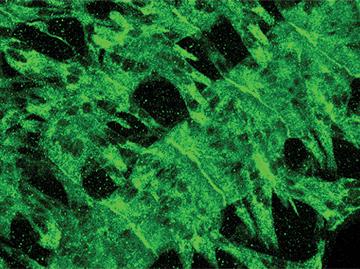
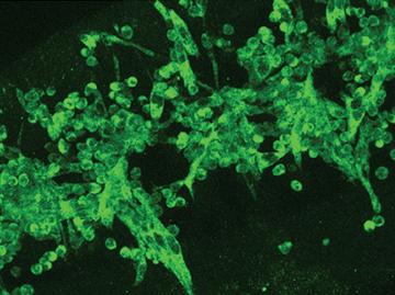
The institute's scientists used genetic methods to remove the gene responsible for the production of the WIP protein from embryos of the fruit fly called "the research Drosophila" (Drosophila). This is how they were able to show that in embryos in which the WIP protein is not formed, normal muscle fibers were not formed. The myoblast cells recognized each other and adhered - but their membranes did not fuse, and they did not form together the multi-nuclear unified structure that makes up the muscle fiber. These findings are published today in the scientific journal Developmental Cell.
This research contributed to a better understanding of the muscle building process, which may help, in the future, to develop advanced ways to heal muscles in humans. One such way may be based on the fusion of stem cells with damaged or degenerated muscle fibers.
Fusion of membranes is an essential step in the creation of different types of bone cells, placental cells and cells of the immune system, as well as in the processes of fertilization and penetration of viruses into living cells. A better understanding of the fusion process may lead to the development of ways to encourage this process in cases where it is desirable, and delay it in cases where it may cause harm.
