An MRI scanner with special software that allows it to monitor the ability of the brain to function. The fMRI is currently used not only for research, but also for the diagnosis of various diseases such as fibromyalgia, for locating specific brain areas before surgery and more
The fMRI scan, an acronym for Functional Magnetic Resonance Imaging (in Hebrew, "functional magnetic resonance imaging"), is a relatively new technology or imaging method, the invention of which brought about a real revolution in the world of research and psychiatry, and some claim that it will become one of the leading research technologies in the future in the following decades.
So what exactly is fMRI?
It is basically an MRI scanner with special software that allows it to monitor the ability of the brain to function. The fMRI is currently used not only for research, but also for diagnosing various diseases such as fibromyalgia, for locating specific brain areas before surgery, and more. In the post below we will explain how the fMRI works and we will also explain the study that presented the famous dead salmon picture - in which they saw brain activity in the fMRI brain of a dead fish (!?!).
how does thefMRI?
The fMRI scan, which was first introduced in 1990, is designed to locate areas that are activated in the brain as a result of performing tasks or as a result of sensory stimulation - for example touching the nose or contracting a muscle express themselves in the brain, and the fMRI is designed to identify the area that is activated in the brain in response to this .
In 1913, a brain surgeon named Wilder Penfield (Wilder Penfield) submitted his doctoral thesis at Princeton University, in which he presented the homunculus, the image of the little man that is drawn inside the brain. This image is used as a clustering of the human body in the brain, and the various organs of the body as reflected in the processing of neural information in the cerebral cortex (while enlarging the areas of the hands, lips and tongue because they get more processing space in the brain). The fMRI makes it possible to actually confirm this research, but in a non-invasive way.
How does he do it?
In order to function, our body cells need a constant supply of oxygen and nutrients, substances that are delivered to them through the blood vessels in our body. There are organs in our body such as the liver and the muscles of the body, which know how to store nutrients (in the form of glycogen) and oxygen (in the form of myoglobin). These stores are designed to provide more oxygen and glucose than the blood vessels can provide, in order to meet the requirements. The brain does not have such stores, so it relies solely on the blood supply for oxygen and nutrients. The blood vessels in the brain (and of course also in the body), have the ability to expand in diameter as a response to certain biochemical changes and thus, if necessary, they know how to supply more oxygen and glucose to the cells in the tissues - in the case of the brain - to the neuron cells.
The MRI scanner is able to measure indirectly the activity of the neurons based on the activity of the regional blood vessels when it is based on the differences in the effects created by the magnetic resonance signal of oxygenated hemoglobin (Oxyhemoglobin) versus the magnetic resonance signal of non-oxygenated hemoglobin (deoxyhemoglobin).
In order for us to better understand the difference in the signal, we will devote a few words to describe the hemoglobin protein.
Hemoglobin is a protein that belongs to a family of proteins called metalloproteins, where its main function is to carry oxygen to the body's cells in the blood system. The name "hemoglobin" contains the two structures that make it up - the "heme" group, a non-proteinaceous molecule consisting of a ring structure in the center of which is an iron atom surrounded by four nitrogen atoms, and the globin protein, a protein in which the "heme" molecule is embedded in a covalent bond. The entire hemoglobin molecule consists of four subunits, which are actually globin units linked to each other, and four "they" groups found within the globin proteins. Each "they" group can bind one oxygen molecule, therefore one hemoglobin molecule has the binding potential of up to four oxygen molecules.
Oxyhemoglobin (oxyhemoglobin) is oxygenated hemoglobin that appears as a bright red substance that is formed when hemoglobin in red blood cells combines with oxygen. This is the form in which the oxygen is transported to the tissues, where it is released. Deoxyhemoglobin (Deoxyhemoglobin) is unoxidized hemoglobin, without oxygen atoms, which is why it has an oval blue color.
How does the MRI scanner manage to differentiate the different signal of oxygenated hemoglobin from non-oxygenated hemoglobin?
The magnetic resonance signal is significantly affected by the number of paired electrons (paired electrons, two electrons that are in the same orbital but have opposite spins) and the number of unpaired electrons (an electron that is alone in an orbital, for this reason the atom or molecule is more reactive to chemical reactions) .
Paired electrons are electrons that do not have a magnetic effect (what is called Diamagnetic) and therefore do not affect the local magnetic resonance signal, while unpaired electrons are magnetized (what is called Paramagnetic), therefore they are able to affect the local magnetic resonance signal, and at high concentrations even turn the area The entire image is dark (in standard spin echo sequences).
Oxyhemoglobin, oxygenated hemoglobin, has no unpaired electrons and therefore has no effect on the local magnetic resonance signal (it is diamagnetic). When it releases its oxygen atoms, it becomes deoxyhemoglobin, deoxygenated hemoglobin, with four unpaired electrons. Now the hemoglobin becomes paramagnetic and the high concentration of deoxyhemoglobin in the blood causes inhomogeneity in the magnetic field and artifacts in the image, elements that impair the strength of the local magnetic resonance signal. This is why oxyhemoglobin appears brighter than deoxyhemoglobin on a T2 MRI image.
But what has been described so far is only half the picture.
As mentioned before, the brain does not have oxygen and energy stores (like for example in the liver and muscles) and therefore it depends solely on the blood supply to it. He has oxyhemoglobin flowing in his cerebral arteries, which, as said, since it is not magnetized, diamagnetic, has no effect on the local magnetic resonance signal. In a normal state, let's say a state of rest, the neurons extract what they need from the blood vessels, leaving in the capillaries mainly the bluish deoxyhemoglobin that has the magnetic effect and then there is a significant decrease in the signal in the activated brain area or shortly after.
However, in the operating state of neurons, meaning when they are activated, the local presence of neurotransmitters causes support cells around them, which are called astrocytes (the large and common cells in the central nervous system, they are shaped like a star and they play many supporting roles in brain tissue and the spinal cord), to release chemicals into The arteries which cause the blood vessels to expand and then provide more oxygen and nutrients to the neurons in the activated area of the brain.
As a result, the increase in blood volume is more than sufficient for the metabolic need that is required, and thus we actually reach a paradoxical situation where there is an increase in the amount of reddish oxyhemoglobin also in the venous capillaries, after their passage through the neurons in the activated brain area. Since oxyhemoglobin is a diamagnetic substance (does not magnetize and does not affect the magnetic resonance signal), there is an increase in the regional magnetic resonance signal relative to the areas of the non-octaminated neurons, which produces a brighter area of magnetic resonance signal in the area of the octaved neurons (or rather, shortly after it, we will reach to that later).
Thus, what happens in practice is that the signal resolution between the activated neurons and the neurons in a resting state (the neurons that are not activated) is based on the relative concentration of oxyhemoglobin in the venous capillaries and therefore this type of imaging is called BOLD - Blood Oxygen Level Dependent functional MRI.
The limitations of BOLD fMRI and on the brain activity of the dead salmon fish
BOLD fMRI technology is a powerful tool in functional research of the brain, but it also has clear limitations due to the way its signal is acquired - these are mainly related to temporal resolution (TE) and spatial accuracy. Temporal resolution refers to the resolution of a measurement with respect to time. Since we indirectly test the activity of the neurons based on the regional increase in the venous capillaries of oxyhemoglobin after the location of the octave neurons, this depends on the orientation of the drainage of the veins that emit the signal, therefore the location of the signal can be detected a few millimeters after the area of the octave neurons.
Another case, relatively famous, that presented to the world the limitation of BOLD fMRI, is related to the background noises by which it can be affected.
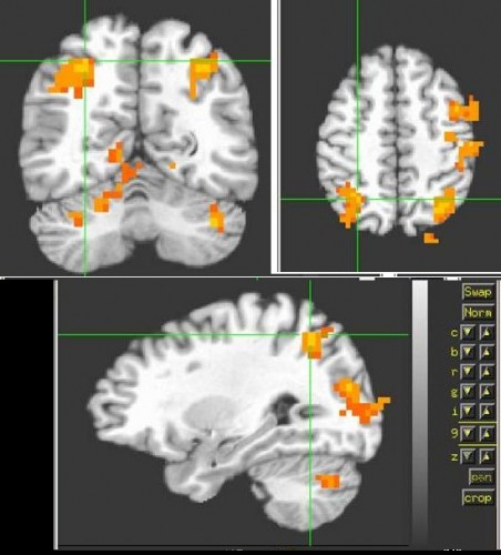
In 2009, Bennett and his associates presented an image, now quite famous, in an article called: Neural correlates of intraspecies perspective-taking in the post mortem Atlantic salmon
The image shows brain activity in a dead salmon in BOLD fMRI in response to the presentation of images of people in different social situations. In at least one image, the researchers were able to demonstrate a significant signal within the salmon's brain, which is supposed to be inactive.
The image and the article that summarized it were not intended to oppose or mock fMRI technology, but to raise awareness of spurious results in fMRI scans, which can be caused by random background noise, if certain statistical corrections are not routinely made when analyzing fMRI results data. The authors of the article attacked the results of many studies that used fMRI, the data of which can be incorrect due to the non-use of correction software for the multiple comparisons problem (a statistical problem caused when several statistical inferences are made at the same time, or when a set of parameters are estimated at the same time based on data sometimes the corrections can also cause problems and reject the null hypothesis, even when it is true). This problem arises specifically from the methodological limitations of using BOLD fMRI.
In conclusion, the fMRI is a fascinating tool that has opened up many options for the world of research. Although it is not without its shortcomings, it is a research tool that undoubtedly has a future ahead of it.
Bibliography and for further reading:
The famous article about the dead salmon fish:
Neural correlates of intraspecies perspective-taking in the post mortem Atlantic salmon
The fMRI and brain imaging portal- https://fmri.co.il/
The Israeli MRI portal- https://mriportal.co.il/
More of the topic in Hayadan:
- It is possible to "watch" brain metabolism using MRI
- Technion researchers have developed new nanometer particles to mark cells for MRI imaging and light microscopy
- The most powerful MRI scanners in the world and what is right for us in the future in the field
The author of the article: Ofer Ben Horin, who has about 20 years of experience in applications, drug research and training in the field of MRI. Author of the bookMRI the complete guide-medicine and physics meetOn the website www.mriguide.co.il
