The blood-brain barrier is a physical barrier of cells that crowd along the walls of blood vessels in the brain, preventing the passage of molecules above a certain size. However, in the pituitary gland, which lies at the base of the brain, there are apparently "broken" blood vessels, which allow large molecules to pass in both directions
The blood-brain barrier is a physical barrier of cells that crowd along the walls of blood vessels in the brain, preventing the passage of molecules above a certain size. However, in the pituitary gland located at the base of the brain, there are apparently "broken" blood vessels that allow large molecules to pass in both directions. Weizmann Institute of Science scientists recently discovered how the blood vessels of the pituitary gland open "windows" to the rest of the body. The research findings, published in the scientific journal Developmental Cell, have possible implications for the treatment of hormonal imbalance or inflammatory diseases of the central nervous system, as well as for the development of future means of introducing drugs into the brain.
"Our research sent us back in time to studies from the beginning of the 20th century on the pituitary gland that laid the foundations for modern neuroendocrinology," says Prof. Gil Lebkowitz from the Department of Molecular Biology of the Cell. "At first it was believed that the pituitary gland is the source of the hormones vasopressin and oxytocin, but in the XNUMXs, Berta and Ernst Scherer, in their research on small freshwater fish, conceived the theory of "neurosecretion". They hypothesized that some brain hormones are produced by nerve cells in an area of the brain called the hypothalamus, and that these hormones are transported along nerve fibers and secreted into the blood vessels in the pituitary gland."
Andopressin and oxytocin were among the first hormones discovered. Vasopressin was first identified as a regulator of kidney function and later of blood pressure; Oxytocin - as a stimulant for uterine contractions during childbirth and milk secretion during breastfeeding. Later it was also discovered that these two hormones have an effect on brain activity, including social behavior, stress and appetite. Andopressin and oxytocin are released from the brain into the body's general circulation through an area at the back of the pituitary gland called the neurohypophysis.
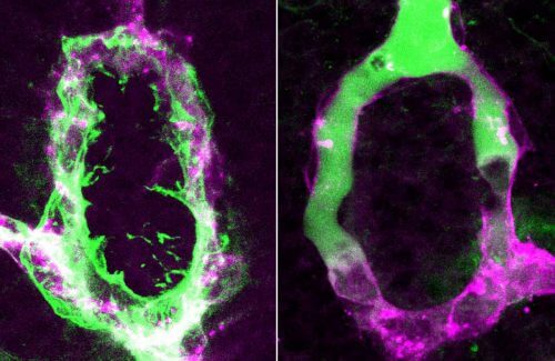
Postdoctoral researcher Dr. Savani Anbalgan and research student Ludmila (Lucy) Gordon from Prof. Lebkowitz's research group focused on the study of the neurohypophysis, where the long extensions (axons) of the nerve cells of the hypothalamus meet blood capillaries where tiny openings are called fenestrae (windows in Latin ). Even after 100 years of research into the development and function of the neurohypophysis, it is still not clear how the capillaries with the "windows" develop, despite the fact that these capillaries branch off from blood vessels, all of which are characterized by the presence of the blood-brain barrier. The researchers in Prof. Lebkowitz's laboratory hypothesized that pituicytes - unique support cells, found only in the neuro-hypophysis, and wrapping the ends of the axons - play a role in the permeability of the local capillaries.
The research team tested the hypothesis in zebrafish embryos, whose transparent bodies allow real-time observation of the neurohypophysis and its unique blood vessels. First, the researchers discovered that the neurohypophysis in fish is almost exactly the same as that in humans. "This is indeed a primitive mechanism of transmitting neuro-hormonal signals between the brain and the body, but it works so well that evolution has not 'touched' it," explains Dr. Anbalagan. Later, the researchers turned to sequencing the genes expressed in the phytocytes, and created transgenic fish, which make it possible to influence some of the molecules they secrete in order to understand the role of each of them.
Prof. Lebkowitz and his colleagues identified two types of communication molecules produced in phytocytes, which make the capillaries permeable. Disruption of these molecules reduced vascular permeability and resulted in the formation of a dense blood-brain barrier. "The windows are dynamic structures," explains Gordon. "The two molecules we identified are active in both the embryo and the adult fish, so that once the "windows" are opened, they will remain open in the future as well." The researchers found that one molecule promotes permeability of the blood vessels by regulating genes that create the "windows", while the other molecule prevents the formation of the blood-brain barrier. "In other words, the body makes sure that the blood vessels are permeable through a double control mechanism," says Prof. Lebkowitz. "It's like walking with both a belt and sneakers."
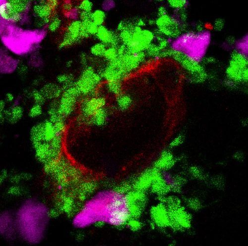
While reviewing the list of molecules secreted by the phytocytes, the researchers noticed the great similarity between this list and the list of molecules secreted in other parts of the brain during inflammation. Such gene expression patterns characterize, for example, autoimmune diseases, neurodegenerative diseases and stroke. Moreover, there is a close connection between the cells that produce molecules that promote inflammation in the protected parts of the brain, and the phytocytes. Both the former and the latter are cells known as glia, support cells that perform many "maintenance" functions in the brain. "We believe that the normal, i.e. the permeable, state of the pituitary gland is similar to a disease state in other parts of the brain, which open their doors only in crisis situations," says Dr. Anbalgan.
Indeed, when the researchers gave the fish an anti-inflammatory drug, they found that gene expression in the phytocytes was blocked and capillary permeability in the neurohypophysis disappeared. This finding provided further evidence that the phytocytes use inflammation-like mechanisms in order to open the "windows" in the capillaries close to them.
Understanding the relationship between the inflammatory response of phytocytes to vascular permeability may advance the understanding of both inflammatory processes in the brain and the normal functioning of the pituitary gland. This may even help to develop new means of delivering essential drugs through the blood-brain barrier.

2 תגובות
Hello again,
I re-read this tab and even the article about the stinging yellow scorpion that can cross the BBB-Blood Brain Barrier by means of a protein found in its venom.
First, I misunderstood the molecules, because if large molecules can pass the blood-brain barrier in the neurohypophysis, it's easy for the small ones to pass.
Second, I still have no idea if I understood this article correctly.
And, if it is theoretically possible to transfer drugs through the small blood capillaries in this area of the brain, to people with neurological and large brain problems, does this mean that it is possible to reach from this area also to the other areas of the brain and heal them?
Also, I didn't understand what the advantages and disadvantages of the two ways are, I meant the possibility of crossing the BBB using animal venom (such as the protein found in scorpion venom) compared to the possibility of crossing the BBB through the neurohypophysis.
Good Morning,
I would like to ask, if possible, whether I understood this article correctly.
To my understanding, glial cells found in the brain (I meant glial cells apart from the phytocytes found only in the neurohypophysis),
Molecules that promote inflammation are produced, which means it is a real inflammation.
Whereas the phytocytes, which are also glial cells but are found only in the neurohypophysis, also secrete molecules that promote inflammation
But it is not a real inflammation.
and in the meeting of the hepatocytes with the small blood capillaries (because they wrap the ends of the axon) instead of causing an inflammatory condition,
They cause the small blood capillaries in the neurohypophysis to open windows, that is, to be permeable to large molecules.
And this is actually a mechanism that bypasses the BBB (at least partially since they manage to make the blood capillaries permeable only to large molecules, the small molecules are still blocked).
Did I understand this article correctly?