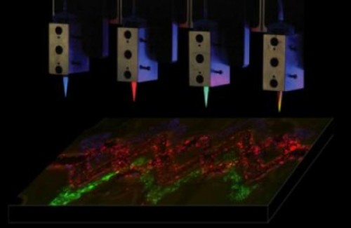
The Harvard researchers are now looking to focus on more realistic functional tissues to screen drugs and test their efficacy and safety. "This is the field where the potential impact of technology will be immediate"

Health and quality of life today depend a lot on science, but nevertheless there are still problems that modern medicine cannot solve, especially when it comes to injuries. Now a new direction is emerging in the emerging field of tissue engineering, which rides on a technology that was developed in general for other needs - XNUMXD printing.
A new method for printing biological tissues was developed at the Wyss Institute for Biologically Inspired Engineering (Wyss Institute for Biologically Inspired Engineering) at Harvard University and the SEAS School of Engineering and Applied Sciences at Harvard, created complex tissues that contain many types of cells and even tiny blood vessels in XNUMXD . According to the researchers, this is an important step towards the long-term goal of tissue engineering: creating realistic enough human tissues to safely and effectively test drugs on.
The method also constitutes an initial but important step towards building functional replacement tissues for injured or disabled tissues that can be designed using a tomography scan, and then printed in XNUMXD at the push of a button for the surgeon to use to repair or replace the damaged tissue.
"This is a fundamental step towards the creation of living tissues in 18D printing," says Jennifer Lewis, the article's lead researcher, a senior faculty member at the Wise Institute, and her partner David Kolasky, a PhD student at SEAS, in a study published on February XNUMX in the journal Advanced Materials.
Researchers in the field of tissue engineering have been trying for years to produce tissues containing human blood vessels strong enough to replace the damaged tissues. Others have previously tried to print human tissue, but they were limited to thin layers of tissue, when the internal cells suffered from a lack of oxygen and nutrients, and there is no effective way to remove carbon dioxide and other waste, so they wither and die.
Nature overcomes the problem by building a network of thin capillaries that supply oxygen and nutrients to the cell and eliminate the waste, so the two decided to imitate this feature. Lewis and her team members had previously recorded a development in the field of XNUMXD printing when they switched from plastics and metals, inert materials, to materials that would allow tissues to only function and not just take the shape of human tissue.
They developed a 'biological ink' - a substance that contains the main components of living tissue. One of the inkheads built the extracellular matrix, the biological material that connects cells to tissues. In addition to the first material, the second ink also contained living cells.
To make blood cells, they needed to develop a third ink with unusual properties: it melts when it cools, instead of when it heats up. This allowed the scientists to first print a connected network of fibers, then melt them by cooling the material and pumping out the liquid to create a network of hollow tubes - the blood vessels.
The Harvard team then developed an approach to test the strength and durability of these blood vessels. They printed three-dimensional tissues in a variety of architectures, and built blood vessels and three types of cells similar to the structure and complexity of solid tissues.
Moreover, when they injected human endothelial cells (cells that line, among other things, the wall of blood cells) into the blood vessel network, these cells regenerated the blood cell network, keeping the cells alive and even growing within the tissue. "Ideally, we want biology to do as much work as possible," Lewis said.
Lewis and her team now want to focus on more realistic functional tissues to screen drugs and test their efficacy and safety. "This is the field where the potential impact of technology will be immediate." Full tissue engineering is still a vision for the future, perhaps another one of those technologies that will bring the sinoglerity?
More of the topic in Hayadan:
